 Movie
Movie Controller


+ Open data

- Basic information
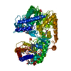
| Entry | Database: PDB / ID: 1dys | ||||||
|---|---|---|---|---|---|---|---|
| Title | Endoglucanase CEL6B from Humicola insolens | ||||||
 Components Components | ENDOGLUCANASE | ||||||
 Keywords Keywords | CELLULASE / HYDROLASE / CELLULOSE DEGRADATION / GLYCOSIDE HYDROLASE FAMILY 6 | ||||||
| Function / homology |  Function and homology information Function and homology information | ||||||
| Biological species |  HUMICOLA INSOLENS (fungus) HUMICOLA INSOLENS (fungus) | ||||||
| Method |  X-RAY DIFFRACTION / MOLECULAR REPLACEMENT / Resolution: 1.6 Å X-RAY DIFFRACTION / MOLECULAR REPLACEMENT / Resolution: 1.6 Å | ||||||
 Authors Authors | Davies, G.J. / Brzozowski, A.M. / Dauter, M. / Varrot, A. / Schulein, M. | ||||||
 Citation Citation | Journal: Biochem.J. / Year: 2000 Title: Structure and Function of Humicola Insolens Family 6 Cellulases: Structure of the Endoglucanase, Cel6B, at 1.6 A Resolution Authors: Davies, G.J. / Brzozowski, A.M. / Dauter, M. / Varrot, A. / Schulein, M. | ||||||
| History |
| ||||||
| Remark 700 | SHEET DETERMINATION METHOD: DSSP THERE ARE SEVERAL BIFURCATED SHEETS IN THIS STRUCTURE. EACH IS ... SHEET DETERMINATION METHOD: DSSP THERE ARE SEVERAL BIFURCATED SHEETS IN THIS STRUCTURE. EACH IS REPRESENTED BY TWO SHEETS WHICH HAVE ONE OR MORE IDENTICAL STRANDS. SHEETS A AND A1 REPRESENT ONE BIFURCATED SHEET. SHEETS C AND C1 REPRESENT ONE BIFURCATED SHEET. |
- Structure visualization
| Structure viewer | Molecule: MolmilJmol/JSmol |
|---|
- Downloads & links
-Download
| PDBx/mmCIF format | 1dys.cif.gz | 159.5 KB | Display | PDBx/mmCIF format |
|---|---|---|---|---|
| PDB format | pdb1dys.ent.gz | 126 KB | Display | PDB format |
| PDBx/mmJSON format | 1dys.json.gz | Tree view | PDBx/mmJSON format | |
| Others |  Other downloads Other downloads |
-Validation report
| Arichive directory | https://data.pdbj.org/pub/pdb/validation_reports/dy/1dysftp://data.pdbj.org/pub/pdb/validation_reports/dy/1dys | HTTPS FTP |
|---|
-Related structure data
| Similar structure data |
|---|


-Links
 PDBj
PDBj- Assembly
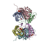
| Deposited unit | 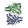
| ||||||||
|---|---|---|---|---|---|---|---|---|---|
| 1 |
| ||||||||
| Unit cell |
| ||||||||
| Noncrystallographic symmetry (NCS) | NCS oper: (Code: given Matrix: (-0.162288, 0.986735, -0.004104), Vector: Details | BIOLOGICAL_UNIT: ACTIVE AS A MONOMER | |
-Components
| #1: Protein | Mass: 37785.832 Da / Num. of mol.: 2 / Fragment: CATALYTIC DOMAIN Source method: isolated from a genetically manipulated source Source: (gene. exp.) HUMICOLA INSOLENS (fungus) / Cellular location: EXCRETED / Production host: #2: Water | ChemComp-HOH / |  Mass: 18.015 Da / Num. of mol.: 901 / Source method: isolated from a natural source / Formula: H2O Mass: 18.015 Da / Num. of mol.: 901 / Source method: isolated from a natural source / Formula: H2OHas protein modification | Y | Sequence details | REFERENCE: SEQUENCE NOT YET DEPOSITED. | |
|---|
-Experimental details
-Experiment
| Experiment | Method: X-RAY DIFFRACTION / Number of used crystals: 1 |
|---|
- Sample preparation
| Crystal | Density Matthews: 2.08 Å3/Da / Density % sol: 40.5 % | ||||||||||||||||||||||||||||||
|---|---|---|---|---|---|---|---|---|---|---|---|---|---|---|---|---|---|---|---|---|---|---|---|---|---|---|---|---|---|---|---|
| Crystal grow | pH: 7.5 Details: THE PROTEIN (20 MG/ML-1) WAS CRYSTALLISED FROM 30% (W/V) PEG 4000K IN 10MM TRIS-ACETATE BUFFER AT PH 7.5 | ||||||||||||||||||||||||||||||
| Crystal grow | *PLUS pH: 8.5 / Method: vapor diffusion, hanging drop | ||||||||||||||||||||||||||||||
| Components of the solutions | *PLUS
|
-Data collection
| Diffraction | Mean temperature: 120 K |
|---|---|
| Diffraction source | Source: ROTATING ANODE / Type: RIGAKU RUH2R / Wavelength: 1.5418 |
| Detector | Type: MARRESEARCH / Detector: IMAGE PLATE / Date: Oct 15, 1997 / Details: LONG MIRRORS (MSC) |
| Radiation | Protocol: SINGLE WAVELENGTH / Monochromatic (M) / Laue (L): M / Scattering type: x-ray |
| Radiation wavelength | Wavelength: 1.5418 Å / Relative weight: 1 |
| Reflection | Resolution: 1.6→15 Å / Num. obs: 78038 / % possible obs: 98.9 % / Redundancy: 3.7 % / Biso Wilson estimate: 21 Å2 / Rmerge(I) obs: 0.065 / Net I/σ(I): 17 |
| Reflection shell | Resolution: 1.6→1.66 Å / Redundancy: 4 % / Rmerge(I) obs: 0.317 / Mean I/σ(I) obs: 3.6 / % possible all: 97.8 |
| Reflection | *PLUS Highest resolution: 1.6 Å / Lowest resolution: 15 Å |
| Reflection shell | *PLUS Highest resolution: 1.6 Å / % possible obs: 98 % |
- Processing
| Software |
| ||||||||||||||||||||||||||||||||||||||||||||||||||||||||||||||||||||||||||||||||||||
|---|---|---|---|---|---|---|---|---|---|---|---|---|---|---|---|---|---|---|---|---|---|---|---|---|---|---|---|---|---|---|---|---|---|---|---|---|---|---|---|---|---|---|---|---|---|---|---|---|---|---|---|---|---|---|---|---|---|---|---|---|---|---|---|---|---|---|---|---|---|---|---|---|---|---|---|---|---|---|---|---|---|---|---|---|---|
| Refinement | Method to determine structure: MOLECULAR REPLACEMENT Starting model: TRICHODERMA REESEI CELLOBIOHYDROLASE II (T A JONES, PERSONAL COMMUNICATION) Resolution: 1.6→15 Å / Cross valid method: THROUGHOUT / σ(F): 0 Details: TWO MOLECULES IN ASYMMETRIC UNIT RESIDUE ALA 182 LIES IN A FORBIDDEN REGION OF THE RAMACHANDRAN PLOT. IT IS PART OF A HYDROGEN-BONDING NETWROK IN THE ACTIVE- SITE AND IS NOT AN ERROR. THE ...Details: TWO MOLECULES IN ASYMMETRIC UNIT RESIDUE ALA 182 LIES IN A FORBIDDEN REGION OF THE RAMACHANDRAN PLOT. IT IS PART OF A HYDROGEN-BONDING NETWROK IN THE ACTIVE- SITE AND IS NOT AN ERROR. THE FIRST TWO RESIDUES ARE NOT VISIBLE IN THE ELECTRON DENSITY. THE STRUCTURE CONTAINS ONLY THE CATALYTIC CORE DOMAIN WHICH TERMINATES WITH RESIDUE ALA 347. THE C-TERMINAL RESIDUE WAS NOT SEEN IN THE DENSITY
| ||||||||||||||||||||||||||||||||||||||||||||||||||||||||||||||||||||||||||||||||||||
| Displacement parameters | Biso mean: 22 Å2 | ||||||||||||||||||||||||||||||||||||||||||||||||||||||||||||||||||||||||||||||||||||
| Refinement step | Cycle: LAST / Resolution: 1.6→15 Å
| ||||||||||||||||||||||||||||||||||||||||||||||||||||||||||||||||||||||||||||||||||||
| Refine LS restraints |
| ||||||||||||||||||||||||||||||||||||||||||||||||||||||||||||||||||||||||||||||||||||
| Software | *PLUS Name: REFMAC / Classification: refinement | ||||||||||||||||||||||||||||||||||||||||||||||||||||||||||||||||||||||||||||||||||||
| Refinement | *PLUS Rfactor obs: 0.18 | ||||||||||||||||||||||||||||||||||||||||||||||||||||||||||||||||||||||||||||||||||||
| Solvent computation | *PLUS | ||||||||||||||||||||||||||||||||||||||||||||||||||||||||||||||||||||||||||||||||||||
| Displacement parameters | *PLUS |
