 Movie
Movie Controller

[English] 日本語
 Yorodumi
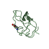
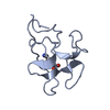
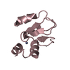
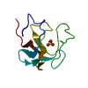
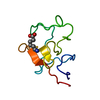
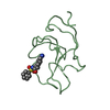
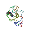
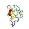
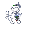
- PDB-1ceb: THE STRUCTURE OF THE NON-COVALENT COMPLEX OF RECOMBINANT KRINGLE ... -
+ Open data

- Basic information
| Entry | Database: PDB / ID: 1ceb | ||||||
|---|---|---|---|---|---|---|---|
| Title | THE STRUCTURE OF THE NON-COVALENT COMPLEX OF RECOMBINANT KRINGLE 1 DOMAIN OF HUMAN PLASMINOGEN WITH AMCHA (TRANS-4-AMINOMETHYLCYCLOHEXANE-1-CARBOXYLIC ACID) | ||||||
 Components Components | PLASMINOGEN | ||||||
 Keywords Keywords | SERINE PROTEASE | ||||||
| Function / homology |  Function and homology information Function and homology informationplasmin / trans-synaptic signaling by BDNF, modulating synaptic transmission / trophoblast giant cell differentiation / tissue remodeling / protein antigen binding / tissue regeneration / mononuclear cell migration / Signaling by PDGF / negative regulation of cell-cell adhesion mediated by cadherin / positive regulation of fibrinolysis ...plasmin / trans-synaptic signaling by BDNF, modulating synaptic transmission / trophoblast giant cell differentiation / tissue remodeling / protein antigen binding / tissue regeneration / mononuclear cell migration / Signaling by PDGF / negative regulation of cell-cell adhesion mediated by cadherin / positive regulation of fibrinolysis / Dissolution of Fibrin Clot / negative regulation of cell-substrate adhesion / myoblast differentiation / biological process involved in interaction with symbiont / labyrinthine layer blood vessel development / muscle cell cellular homeostasis / Activation of Matrix Metalloproteinases / apolipoprotein binding / extracellular matrix disassembly / positive regulation of blood vessel endothelial cell migration / negative regulation of fibrinolysis / fibrinolysis / Degradation of the extracellular matrix / serine-type peptidase activity / platelet alpha granule lumen / kinase binding / Schaffer collateral - CA1 synapse / Regulation of Insulin-like Growth Factor (IGF) transport and uptake by Insulin-like Growth Factor Binding Proteins (IGFBPs) / blood coagulation / Platelet degranulation / protein-folding chaperone binding / endopeptidase activity / protease binding / collagen-containing extracellular matrix / blood microparticle / protein domain specific binding / negative regulation of cell population proliferation / external side of plasma membrane / serine-type endopeptidase activity / signaling receptor binding / glutamatergic synapse / enzyme binding / cell surface / proteolysis / extracellular space / extracellular exosome / extracellular region / plasma membrane Similarity search - Function | ||||||
| Biological species |  Homo sapiens (human) Homo sapiens (human) | ||||||
| Method |  X-RAY DIFFRACTION / Resolution: 2.07 Å X-RAY DIFFRACTION / Resolution: 2.07 Å | ||||||
 Authors Authors | Tulinsky, A. / Mathews, I.I. | ||||||
 Citation Citation | Journal: Biochemistry / Year: 1996 Title: Crystal structures of the recombinant kringle 1 domain of human plasminogen in complexes with the ligands epsilon-aminocaproic acid and trans-4-(aminomethyl)cyclohexane-1-carboxylic Acid. Authors: Mathews, I.I. / Vanderhoff-Hanaver, P. / Castellino, F.J. / Tulinsky, A. #1: Journal: Eur.J.Biochem. / Year: 1994Title: 1H-NMR Assignments and Secondary Structure of Human Plasminogen Kringle 1 Authors: Rejante, M.R. / Llinas, M. #2: Journal: Blood Coagulation Fibrinolysis / Year: 1994Title: The Structure of Recombinant Plasminogen Kringle 1 and the Fibrin Binding Site Authors: Wu, T.-P. / Padmanabhan, K.P. / Tulinsky, A. #3: Journal: Proteins / Year: 1988Title: Lysine(Slash)Fibrin Binding Sites of Kringles Modeled After the Structure of Kringle 1 of Prothrombin Authors: Tulinsky, A. / Park, C.H. / Mao, B. / Llinas, M. | ||||||
| History |
|
- Structure visualization
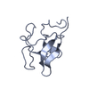
| Structure viewer | Molecule: MolmilJmol/JSmol |
|---|
- Downloads & links
-Download
| PDBx/mmCIF format | 1ceb.cif.gz | 45.8 KB | Display | PDBx/mmCIF format |
|---|---|---|---|---|
| PDB format | pdb1ceb.ent.gz | 35.5 KB | Display | PDB format |
| PDBx/mmJSON format | 1ceb.json.gz | Tree view | PDBx/mmJSON format | |
| Others |  Other downloads Other downloads |
-Validation report
| Summary document | 1ceb_validation.pdf.gz | 393.8 KB | Display | wwPDB validaton report |
|---|---|---|---|---|
| Full document | 1ceb_full_validation.pdf.gz | 417 KB | Display | |
| Data in XML | 1ceb_validation.xml.gz | 9.3 KB | Display | |
| Data in CIF | 1ceb_validation.cif.gz | 13.3 KB | Display | |
| Arichive directory | https://data.pdbj.org/pub/pdb/validation_reports/ce/1cebftp://data.pdbj.org/pub/pdb/validation_reports/ce/1ceb | HTTPS FTP |
-Related structure data
-Links
 PDBj
PDBj


- Assembly
| Deposited unit | 
| ||||||||
|---|---|---|---|---|---|---|---|---|---|
| 1 | 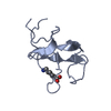
| ||||||||
| 2 | 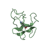
| ||||||||
| Unit cell |
| ||||||||
| Atom site foot note | 1: CIS PROLINE - PRO A 30 / 2: CIS PROLINE - PRO B 30 | ||||||||
| Noncrystallographic symmetry (NCS) | NCS oper: (Code: given Matrix: (0.99956, -0.02947, 0.003414), Vector: |
-Components
| #1: Protein | Mass: 10166.165 Da / Num. of mol.: 2 / Fragment: KRINGLE 1 Source method: isolated from a genetically manipulated source Source: (gene. exp.) Homo sapiens (human) / Organ: BLOOD / Production host:  #2: Chemical | 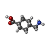  Mass: 157.210 Da / Num. of mol.: 2 / Source method: obtained synthetically / Formula: C8H15NO2 / Comment: medication*YM Mass: 157.210 Da / Num. of mol.: 2 / Source method: obtained synthetically / Formula: C8H15NO2 / Comment: medication*YM#3: Water | ChemComp-HOH / |  Mass: 18.015 Da / Num. of mol.: 150 / Source method: isolated from a natural source / Formula: H2O Mass: 18.015 Da / Num. of mol.: 150 / Source method: isolated from a natural source / Formula: H2O |
|---|
-Experimental details
-Experiment
| Experiment | Method: X-RAY DIFFRACTION |
|---|
- Sample preparation
| Crystal | Density Matthews: 1.91 Å3/Da / Density % sol: 35.65 % | |||||||||||||||
|---|---|---|---|---|---|---|---|---|---|---|---|---|---|---|---|---|
| Crystal | *PLUS Density % sol: 37 % | |||||||||||||||
| Crystal grow | *PLUS pH: 6.5 / Method: vapor diffusion, hanging drop / Details: seeding | |||||||||||||||
| Components of the solutions | *PLUS
|
-Data collection
| Diffraction | Mean temperature: 295 K |
|---|---|
| Diffraction source | Source: ROTATING ANODE / Type: RIGAKU RU200 / Wavelength: 1.5418 |
| Detector | Type: RIGAKU RAXIS IIC / Detector: IMAGE PLATE |
| Radiation | Monochromatic (M) / Laue (L): M / Scattering type: x-ray |
| Radiation wavelength | Wavelength: 1.5418 Å / Relative weight: 1 |
| Reflection | Highest resolution: 2.06 Å / Num. obs: 7028 / % possible obs: 70 % / Observed criterion σ(I): 1 / Redundancy: 2.6 % / Rmerge(I) obs: 0.0795 / Net I/σ(I): 1 |
| Reflection shell | Resolution: 2.07→2.25 Å / Rmerge(I) obs: 0.14 / Mean I/σ(I) obs: 1 / % possible all: 56 |
| Reflection | *PLUS Highest resolution: 2.07 Å / Lowest resolution: 10 Å / Num. measured all: 17924 |
| Reflection shell | *PLUS Highest resolution: 2.07 Å / Lowest resolution: 2.5 Å / % possible obs: 60 % / Rmerge(I) obs: 0.133 |
- Processing
| Software |
| ||||||||||||||||||||||||||||||||||||||||||||||||||||||||||||||||||||||||||||||||||||
|---|---|---|---|---|---|---|---|---|---|---|---|---|---|---|---|---|---|---|---|---|---|---|---|---|---|---|---|---|---|---|---|---|---|---|---|---|---|---|---|---|---|---|---|---|---|---|---|---|---|---|---|---|---|---|---|---|---|---|---|---|---|---|---|---|---|---|---|---|---|---|---|---|---|---|---|---|---|---|---|---|---|---|---|---|---|
| Refinement | Resolution: 2.07→7 Å / σ(F): 3 Details: NO ELECTRON DENSITY WAS OBSERVED FOR THE INTERKRINGLE RESIDUES 3A - 2A AND 80 - 86 IN BOTH MOLECULES.
| ||||||||||||||||||||||||||||||||||||||||||||||||||||||||||||||||||||||||||||||||||||
| Refinement step | Cycle: LAST / Resolution: 2.07→7 Å
| ||||||||||||||||||||||||||||||||||||||||||||||||||||||||||||||||||||||||||||||||||||
| Refine LS restraints |
|
