 Movie
Movie Controller

[English] 日本語
 Yorodumi
- PDB-1c82: MECHANISM OF HYALURONAN BINDING AND DEGRADATION: STRUCTURE OF STR... -
+ Open data

- Basic information
| Entry | Database: PDB / ID: 1c82 | |||||||||
|---|---|---|---|---|---|---|---|---|---|---|
| Title | MECHANISM OF HYALURONAN BINDING AND DEGRADATION: STRUCTURE OF STREPTOCOCCUS PNEUMONIAE HYALURONATE LYASE IN COMPLEX WITH HYALURONIC ACID DISACCHARIDE AT 1.7 A RESOLUTION | |||||||||
 Components Components | HYALURONATE LYASE | |||||||||
 Keywords Keywords | LYASE / PROTEIN-CARBOHYDRATE COMPLEX | |||||||||
| Function / homology |  Function and homology information Function and homology informationhyaluronate lyase / hyaluronate lyase activity / carbohydrate binding / carbohydrate metabolic process / extracellular region Similarity search - Function | |||||||||
| Biological species |   Streptococcus pneumoniae (bacteria) Streptococcus pneumoniae (bacteria) | |||||||||
| Method |  X-RAY DIFFRACTION / SYNCHROTRON / Resolution: 1.7 Å X-RAY DIFFRACTION / SYNCHROTRON / Resolution: 1.7 Å | |||||||||
 Authors Authors | Ponnuraj, K. / Jedrzejas, M.J. | |||||||||
 Citation Citation | Journal: J.Mol.Biol. / Year: 2000 Title: Mechanism of hyaluronan binding and degradation: structure of Streptococcus pneumoniae hyaluronate lyase in complex with hyaluronic acid disaccharide at 1.7 A resolution. Authors: Ponnuraj, K. / Jedrzejas, M.J. #1: Journal: Embo J. / Year: 2000Title: Structural Basis of Hyaluronan Degradation by Streptococcus pneumoniae Hyaluronate Lyase Authors: Li, S. / Kelly, S.J. / Lamani, E. / Ferraroni, M. / Jedrzejas, M.J. | |||||||||
| History |
| |||||||||
| Remark 999 | SEQUENCE THE SIX RESIDUE HIS TAG WAS NOT SEEN IN THE DENSITY. THE ELECTRON DENSITY OF RESIDUE 731 ...SEQUENCE THE SIX RESIDUE HIS TAG WAS NOT SEEN IN THE DENSITY. THE ELECTRON DENSITY OF RESIDUE 731 SUGGESTS IT IS A VALINE. |
- Structure visualization
| Structure viewer | Molecule: MolmilJmol/JSmol |
|---|
- Downloads & links
-Download
| PDBx/mmCIF format | 1c82.cif.gz | 172.6 KB | Display | PDBx/mmCIF format |
|---|---|---|---|---|
| PDB format | pdb1c82.ent.gz | 131.8 KB | Display | PDB format |
| PDBx/mmJSON format | 1c82.json.gz | Tree view | PDBx/mmJSON format | |
| Others |  Other downloads Other downloads |
-Validation report
| Arichive directory | https://data.pdbj.org/pub/pdb/validation_reports/c8/1c82ftp://data.pdbj.org/pub/pdb/validation_reports/c8/1c82 | HTTPS FTP |
|---|
-Related structure data
| Similar structure data |
|---|


-Links
 PDBj
PDBj


- Assembly
| Deposited unit | 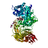
| ||||||||
|---|---|---|---|---|---|---|---|---|---|
| 1 |
| ||||||||
| Unit cell |
|
-Components
| #1: Protein | Mass: 83561.938 Da / Num. of mol.: 1 Source method: isolated from a genetically manipulated source Source: (gene. exp.) Streptococcus pneumoniae (bacteria) / Description: ESCHERICHIA COLI / Species (production host): Escherichia coli / Production host: | ||||||
|---|---|---|---|---|---|---|---|
| #2: Polysaccharide | Source method: isolated from a genetically manipulated source #3: Chemical |   Mass: 136.989 Da / Num. of mol.: 2 / Source method: obtained synthetically / Formula: C2H6AsO2 Mass: 136.989 Da / Num. of mol.: 2 / Source method: obtained synthetically / Formula: C2H6AsO2#4: Chemical |   Mass: 22.990 Da / Num. of mol.: 2 / Source method: obtained synthetically / Formula: Na Mass: 22.990 Da / Num. of mol.: 2 / Source method: obtained synthetically / Formula: Na#5: Water | ChemComp-HOH / |  Mass: 18.015 Da / Num. of mol.: 573 / Source method: isolated from a natural source / Formula: H2O Mass: 18.015 Da / Num. of mol.: 573 / Source method: isolated from a natural source / Formula: H2O |
-Experimental details
-Experiment
| Experiment | Method: X-RAY DIFFRACTION / Number of used crystals: 1 |
|---|
- Sample preparation
| Crystal | Density Matthews: 2.6 Å3/Da / Density % sol: 52.7 % | ||||||||||||||||||||
|---|---|---|---|---|---|---|---|---|---|---|---|---|---|---|---|---|---|---|---|---|---|
| Crystal grow | Temperature: 293 K / Method: evaporation / pH: 6 Details: AMMONIUM SULFATE, SODIUM CACODYLATE, pH 6.0, EVAPORATION, temperature 293K | ||||||||||||||||||||
| Crystal grow | *PLUS Method: vapor diffusion, hanging drop | ||||||||||||||||||||
| Components of the solutions | *PLUS
|
-Data collection
| Diffraction | Mean temperature: 100 K |
|---|---|
| Diffraction source | Source: SYNCHROTRON / Site: APS  / Beamline: 19-ID / Wavelength: 0.9793 / Beamline: 19-ID / Wavelength: 0.9793 |
| Detector | Type: OXFORD / Detector: CCD |
| Radiation | Protocol: SINGLE WAVELENGTH / Monochromatic (M) / Laue (L): M / Scattering type: x-ray |
| Radiation wavelength | Wavelength: 0.9793 Å / Relative weight: 1 |
| Reflection | Resolution: 1.7→100 Å / Num. all: 495691 / Num. obs: 95261 / % possible obs: 98.5 % / Redundancy: 5.2 % / Rmerge(I) obs: 0.055 / Net I/σ(I): 23.4 |
| Reflection shell | Resolution: 1.7→1.76 Å / Redundancy: 3.9 % / Rmerge(I) obs: 0.336 / Num. unique all: 9367 / % possible all: 98 |
| Reflection | *PLUS Num. measured all: 495691 |
| Reflection shell | *PLUS % possible obs: 98 % / Mean I/σ(I) obs: 4.9 |
- Processing
| Software |
| ||||||||||||||||||||
|---|---|---|---|---|---|---|---|---|---|---|---|---|---|---|---|---|---|---|---|---|---|
| Refinement | Resolution: 1.7→40 Å / σ(F): 2 / Stereochemistry target values: Engh & Huber
| ||||||||||||||||||||
| Refinement step | Cycle: LAST / Resolution: 1.7→40 Å
| ||||||||||||||||||||
| Refine LS restraints | Type: x_bond_d / Dev ideal: 0.009 | ||||||||||||||||||||
| Software | *PLUS Name: X-PLOR / Version: 3.85 / Classification: refinement | ||||||||||||||||||||
| Refine LS restraints | *PLUS
|
