 Movie
Movie Controller

[English] 日本語
 Yorodumi
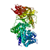
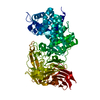
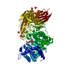
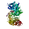
- PDB-1ojo: SPECIFICITY AND MECHANISM OF STREPTOCOCCUS PNEUMONIAE HYALURONATE... -
+ Open data

- Basic information
| Entry | Database: PDB / ID: 1ojo | |||||||||
|---|---|---|---|---|---|---|---|---|---|---|
| Title | SPECIFICITY AND MECHANISM OF STREPTOCOCCUS PNEUMONIAE HYALURONATE LYASE: COMPLEX OF THE TYR408PHE MUTANT WITH 4-SULPHATED CHONDROITIN DISACCHARIDE | |||||||||
 Components Components | HYALURONATE LYASE | |||||||||
 Keywords Keywords | LYASE / PROTEIN-CARBOHYDRATE COMPLEX | |||||||||
| Function / homology |  Function and homology information Function and homology informationhyaluronate lyase / hyaluronate lyase activity / carbohydrate binding / carbohydrate metabolic process / extracellular region Similarity search - Function | |||||||||
| Biological species |   STREPTOCOCCUS PNEUMONIAE (bacteria) STREPTOCOCCUS PNEUMONIAE (bacteria) | |||||||||
| Method |  X-RAY DIFFRACTION / SYNCHROTRON / MOLECULAR REPLACEMENT / Resolution: 1.75 Å X-RAY DIFFRACTION / SYNCHROTRON / MOLECULAR REPLACEMENT / Resolution: 1.75 Å | |||||||||
 Authors Authors | Rigden, D.J. / Jedrzejas, M.J. | |||||||||
 Citation Citation | Journal: J.Biol.Chem. / Year: 2003 Title: Structures of Streptococcus Pneumoniae Hyaluronate Lyase in Complex with Chondroitin and Chondroitin Sulfate Disaccharides: Insights Into Specificity and Mechanism of Action Authors: Rigden, D.J. / Jedrzejas, M.J. #1: Journal: Embo J. / Year: 2000Title: Structural Basis of Hyaluronan Degradation by Streptococcus Pneumoniae Hyaluronate Lyase Authors: Li, S. / Kelly, S.J. / Lamani, E. / Ferraroni, M. / Jedrzejas, M.J. | |||||||||
| History |
| |||||||||
| Remark 700 | SHEET THE SHEET STRUCTURE OF THIS MOLECULE IS BIFURCATED. IN ORDER TO REPRESENT THIS FEATURE IN ... SHEET THE SHEET STRUCTURE OF THIS MOLECULE IS BIFURCATED. IN ORDER TO REPRESENT THIS FEATURE IN THE SHEET RECORDS BELOW, TWO SHEETS ARE DEFINED. |
- Structure visualization
| Structure viewer | Molecule: MolmilJmol/JSmol |
|---|
- Downloads & links
-Download
| PDBx/mmCIF format | 1ojo.cif.gz | 175.6 KB | Display | PDBx/mmCIF format |
|---|---|---|---|---|
| PDB format | pdb1ojo.ent.gz | 135.8 KB | Display | PDB format |
| PDBx/mmJSON format | 1ojo.json.gz | Tree view | PDBx/mmJSON format | |
| Others |  Other downloads Other downloads |
-Validation report
| Arichive directory | https://data.pdbj.org/pub/pdb/validation_reports/oj/1ojoftp://data.pdbj.org/pub/pdb/validation_reports/oj/1ojo | HTTPS FTP |
|---|
-Related structure data
| Related structure data | 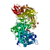 1ojmC  1ojnC  1ojpC  1c82S S: Starting model for refinement C: citing same article ( |
|---|---|
| Similar structure data |


-Links
 PDBj
PDBj


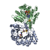
- Assembly
| Deposited unit | 
| ||||||||
|---|---|---|---|---|---|---|---|---|---|
| 1 |
| ||||||||
| Unit cell |
|
-Components
| #1: Protein | Mass: 83646.078 Da / Num. of mol.: 1 / Fragment: HYALURONATE LYASE, RESIDUES 285-1009 / Mutation: YES Source method: isolated from a genetically manipulated source Source: (gene. exp.) STREPTOCOCCUS PNEUMONIAE (bacteria) / Production host: | ||||||
|---|---|---|---|---|---|---|---|
| #2: Polysaccharide | 4-deoxy-alpha-L-threo-hex-4-enopyranuronic acid-(1-3)-2-acetamido-2-deoxy-4-O-sulfo-beta-D-galactopyranose Source method: isolated from a genetically manipulated source | ||||||
| #3: Chemical | ChemComp-SO4 /   Mass: 96.063 Da / Num. of mol.: 4 / Source method: obtained synthetically / Formula: SO4 Mass: 96.063 Da / Num. of mol.: 4 / Source method: obtained synthetically / Formula: SO4#4: Water | ChemComp-HOH / |  Mass: 18.015 Da / Num. of mol.: 536 / Source method: isolated from a natural source / Formula: H2O Mass: 18.015 Da / Num. of mol.: 536 / Source method: isolated from a natural source / Formula: H2OCompound details | ENGINEERED | Sequence details | THE PROTEIN USED WAS THE Y408F MUTANT. THE SIX RESIDUE HIS TAG, ALONG WITH RESIDUES 168-169 AND ...THE PROTEIN USED WAS THE Y408F MUTANT. THE SIX RESIDUE HIS TAG, ALONG WITH RESIDUES 168-169 AND 892, WERE NOT SEEN IN THE DENSITY. THE ELECTRON DENSITY OF RESIDUE 731 SUGGESTS IT IS A VALINE THE SEQUENCE CONFLICT AT RESIDUE A196 (SWS RESIDUE 313) IS MARKED AS A KNOWN SEQUENCE CONFLICT IN THE SWS ENTRY. | |
-Experimental details
-Experiment
| Experiment | Method: X-RAY DIFFRACTION / Number of used crystals: 1 |
|---|
- Sample preparation
| Crystal | Density Matthews: 3.05 Å3/Da / Density % sol: 59.39 % | ||||||||||||||||||||||||||||||||||||||||||
|---|---|---|---|---|---|---|---|---|---|---|---|---|---|---|---|---|---|---|---|---|---|---|---|---|---|---|---|---|---|---|---|---|---|---|---|---|---|---|---|---|---|---|---|
| Crystal grow | pH: 6 Details: 3.5M AMMONIUM SULFATE, 200MM SODIUM CACODYLATE, PH 6.0 | ||||||||||||||||||||||||||||||||||||||||||
| Crystal grow | *PLUS pH: 7.4 / Method: vapor diffusion, hanging drop | ||||||||||||||||||||||||||||||||||||||||||
| Components of the solutions | *PLUS
|
-Data collection
| Diffraction | Mean temperature: 100 K |
|---|---|
| Diffraction source | Source: SYNCHROTRON / Site: APS  / Beamline: 19-ID / Wavelength: 0.9793 / Beamline: 19-ID / Wavelength: 0.9793 |
| Detector | Type: OXFORD / Detector: CCD / Date: Jun 15, 2002 |
| Radiation | Protocol: SINGLE WAVELENGTH / Monochromatic (M) / Laue (L): M / Scattering type: x-ray |
| Radiation wavelength | Wavelength: 0.9793 Å / Relative weight: 1 |
| Reflection | Resolution: 1.5→39.96 Å / Num. obs: 63869 / % possible obs: 86.7 % / Redundancy: 7.9 % / Biso Wilson estimate: 19.2 Å2 / Rmerge(I) obs: 0.089 / Net I/σ(I): 25 |
| Reflection shell | Resolution: 1.75→1.81 Å / Redundancy: 7.4 % / Rmerge(I) obs: 0.757 / Mean I/σ(I) obs: 2.2 / % possible all: 100 |
| Reflection | *PLUS Highest resolution: 1.75 Å / % possible obs: 100 % / Redundancy: 7.9 % / Rmerge(I) obs: 0.089 |
| Reflection shell | *PLUS % possible obs: 100 % / Redundancy: 7.4 % / Rmerge(I) obs: 0.757 / Mean I/σ(I) obs: 2.2 |
- Processing
| Software |
| ||||||||||||||||||||||||||||||||||||||||||||||||||||||||||||||||||||||||||||||||
|---|---|---|---|---|---|---|---|---|---|---|---|---|---|---|---|---|---|---|---|---|---|---|---|---|---|---|---|---|---|---|---|---|---|---|---|---|---|---|---|---|---|---|---|---|---|---|---|---|---|---|---|---|---|---|---|---|---|---|---|---|---|---|---|---|---|---|---|---|---|---|---|---|---|---|---|---|---|---|---|---|---|
| Refinement | Method to determine structure: MOLECULAR REPLACEMENT Starting model: PDB ENTRY 1C82 Resolution: 1.75→39.96 Å / Rfactor Rfree error: 0.003 / Data cutoff high absF: 2256586 / Cross valid method: THROUGHOUT / σ(F): 0
| ||||||||||||||||||||||||||||||||||||||||||||||||||||||||||||||||||||||||||||||||
| Solvent computation | Solvent model: FLAT MODEL / Bsol: 49.7568 Å2 / ksol: 0.400055 e/Å3 | ||||||||||||||||||||||||||||||||||||||||||||||||||||||||||||||||||||||||||||||||
| Displacement parameters | Biso mean: 25.2 Å2
| ||||||||||||||||||||||||||||||||||||||||||||||||||||||||||||||||||||||||||||||||
| Refine analyze |
| ||||||||||||||||||||||||||||||||||||||||||||||||||||||||||||||||||||||||||||||||
| Refinement step | Cycle: LAST / Resolution: 1.75→39.96 Å
| ||||||||||||||||||||||||||||||||||||||||||||||||||||||||||||||||||||||||||||||||
| Refine LS restraints |
| ||||||||||||||||||||||||||||||||||||||||||||||||||||||||||||||||||||||||||||||||
| LS refinement shell | Resolution: 1.75→1.81 Å / Rfactor Rfree error: 0.015 / Total num. of bins used: 10
| ||||||||||||||||||||||||||||||||||||||||||||||||||||||||||||||||||||||||||||||||
| Xplor file |
| ||||||||||||||||||||||||||||||||||||||||||||||||||||||||||||||||||||||||||||||||
| Refinement | *PLUS Num. reflection obs: 90026 | ||||||||||||||||||||||||||||||||||||||||||||||||||||||||||||||||||||||||||||||||
| Solvent computation | *PLUS | ||||||||||||||||||||||||||||||||||||||||||||||||||||||||||||||||||||||||||||||||
| Displacement parameters | *PLUS | ||||||||||||||||||||||||||||||||||||||||||||||||||||||||||||||||||||||||||||||||
| Refine LS restraints | *PLUS
|
