 Movie
Movie Controller


+ Open data

- Basic information
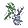
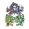
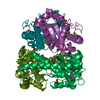
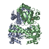
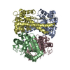
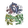
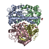
| Entry | Database: PDB / ID: 1bs3 | ||||||
|---|---|---|---|---|---|---|---|
| Title | P.SHERMANII SOD(FE+3) FLUORIDE | ||||||
 Components Components | SUPEROXIDE DISMUTASE | ||||||
 Keywords Keywords | OXIDOREDUCTASE / SUPEROXIDE DISMUTASE / FLUORIDE | ||||||
| Function / homology |  Function and homology information Function and homology informationsuperoxide dismutase / superoxide dismutase activity / metal ion binding Similarity search - Function | ||||||
| Biological species |  Propionibacterium freudenreichii subsp. shermanii (bacteria) Propionibacterium freudenreichii subsp. shermanii (bacteria) | ||||||
| Method |  X-RAY DIFFRACTION / Resolution: 1.55 Å X-RAY DIFFRACTION / Resolution: 1.55 Å | ||||||
 Authors Authors | Schmidt, M. | ||||||
 Citation Citation | Journal: Eur.J.Biochem. / Year: 1999 Title: Manipulating the coordination mumber of the ferric iron within the cambialistic superoxide dismutase of Propionibacterium shermanii by changing the pH-value A crystallographic analysis Authors: Schmidt, M. | ||||||
| History |
|
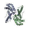
- Structure visualization
| Structure viewer | Molecule: MolmilJmol/JSmol |
|---|
- Downloads & links
-Download
| PDBx/mmCIF format | 1bs3.cif.gz | 101 KB | Display | PDBx/mmCIF format |
|---|---|---|---|---|
| PDB format | pdb1bs3.ent.gz | 77.9 KB | Display | PDB format |
| PDBx/mmJSON format | 1bs3.json.gz | Tree view | PDBx/mmJSON format | |
| Others |  Other downloads Other downloads |
-Validation report
| Arichive directory | https://data.pdbj.org/pub/pdb/validation_reports/bs/1bs3ftp://data.pdbj.org/pub/pdb/validation_reports/bs/1bs3 | HTTPS FTP |
|---|
-Related structure data
| Related structure data | 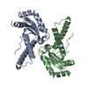 1bsmC  1bt8C 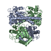 1ar5S S: Starting model for refinement C: citing same article ( |
|---|---|
| Similar structure data |


-Links
 PDBj
PDBj
- Assembly
| Deposited unit | 
| ||||||||
|---|---|---|---|---|---|---|---|---|---|
| 1 | 
| ||||||||
| Unit cell |
| ||||||||
| Noncrystallographic symmetry (NCS) | NCS oper: (Code: given Matrix: (0.2028, -0.0019, -0.9792), Vector: |
-Components
| #1: Protein | Mass: 22659.326 Da / Num. of mol.: 2 / Source method: isolated from a natural source Details: COMPETITIVELY INHIBITED BY FLOURIDE. FLUORIDE OCCUPIES SUBSTRATE BINDING SITE. Source: (natural) Propionibacterium freudenreichii subsp. shermanii (bacteria)Cellular location: CYTOPLASM / Species: Propionibacterium freudenreichii / Strain: PZ3 / References: UniProt: P80293, superoxide dismutase #2: Chemical |   Mass: 55.845 Da / Num. of mol.: 2 / Source method: obtained synthetically / Formula: Fe Mass: 55.845 Da / Num. of mol.: 2 / Source method: obtained synthetically / Formula: Fe#3: Chemical |   Mass: 18.998 Da / Num. of mol.: 2 / Source method: obtained synthetically / Formula: F Mass: 18.998 Da / Num. of mol.: 2 / Source method: obtained synthetically / Formula: F#4: Water | ChemComp-HOH / |  Mass: 18.015 Da / Num. of mol.: 478 / Source method: isolated from a natural source / Formula: H2O Mass: 18.015 Da / Num. of mol.: 478 / Source method: isolated from a natural source / Formula: H2O |
|---|
-Experimental details
-Experiment
| Experiment | Method: X-RAY DIFFRACTION / Number of used crystals: 1 |
|---|
- Sample preparation
| Crystal | Density Matthews: 2 Å3/Da / Density % sol: 32 % | |||||||||||||||||||||||||
|---|---|---|---|---|---|---|---|---|---|---|---|---|---|---|---|---|---|---|---|---|---|---|---|---|---|---|
| Crystal grow | pH: 6.1 / Details: pH 6.1 | |||||||||||||||||||||||||
| Crystal grow | *PLUS Temperature: 4 ℃ / Method: vapor diffusion, hanging drop | |||||||||||||||||||||||||
| Components of the solutions | *PLUS
|
-Data collection
| Diffraction | Mean temperature: 295 K |
|---|---|
| Diffraction source | Source: ROTATING ANODE / Type: ENRAF-NONIUS FR591 / Wavelength: 1.5418 |
| Detector | Type: SIEMENS / Detector: AREA DETECTOR / Date: Sep 1, 1997 |
| Radiation | Monochromator: GRAPHITE(002) / Monochromatic (M) / Laue (L): M / Scattering type: x-ray |
| Radiation wavelength | Wavelength: 1.5418 Å / Relative weight: 1 |
| Reflection | Resolution: 1.55→39.7 Å / Num. obs: 45799 / % possible obs: 83.1 % / Observed criterion σ(I): 2 / Redundancy: 5.6 % / Biso Wilson estimate: 13.7 Å2 / Rsym value: 0.034 / Net I/σ(I): 18 |
| Reflection shell | Resolution: 1.55→1.62 Å / Redundancy: 3.6 % / Mean I/σ(I) obs: 7.6 / Rsym value: 0.148 / % possible all: 67 |
| Reflection | *PLUS Num. obs: 45923 / % possible obs: 78 % / Redundancy: 5.1 % / Rmerge(I) obs: 0.036 |
- Processing
| Software |
| ||||||||||||||||||||||||||||||||||||||||||||||||||||||||||||||||||||||||||||||||
|---|---|---|---|---|---|---|---|---|---|---|---|---|---|---|---|---|---|---|---|---|---|---|---|---|---|---|---|---|---|---|---|---|---|---|---|---|---|---|---|---|---|---|---|---|---|---|---|---|---|---|---|---|---|---|---|---|---|---|---|---|---|---|---|---|---|---|---|---|---|---|---|---|---|---|---|---|---|---|---|---|---|
| Refinement | Starting model: PDB ENTRY 1AR5 Resolution: 1.55→9 Å / Isotropic thermal model: RESTRAINED / Cross valid method: THROUGHOUT / σ(F): 3.5 / Details: IRON AND FLUORIDE REFINED UNRESTRAINED
| ||||||||||||||||||||||||||||||||||||||||||||||||||||||||||||||||||||||||||||||||
| Displacement parameters | Biso mean: 14.1 Å2 | ||||||||||||||||||||||||||||||||||||||||||||||||||||||||||||||||||||||||||||||||
| Refine analyze | Luzzati d res low obs: 5 Å / Luzzati sigma a obs: 0.15 Å | ||||||||||||||||||||||||||||||||||||||||||||||||||||||||||||||||||||||||||||||||
| Refinement step | Cycle: LAST / Resolution: 1.55→9 Å
| ||||||||||||||||||||||||||||||||||||||||||||||||||||||||||||||||||||||||||||||||
| Refine LS restraints |
| ||||||||||||||||||||||||||||||||||||||||||||||||||||||||||||||||||||||||||||||||
| LS refinement shell | Resolution: 1.55→1.62 Å / Total num. of bins used: 8
| ||||||||||||||||||||||||||||||||||||||||||||||||||||||||||||||||||||||||||||||||
| Xplor file |
| ||||||||||||||||||||||||||||||||||||||||||||||||||||||||||||||||||||||||||||||||
| Software | *PLUS Name: X-PLOR / Version: 3.851 / Classification: refinement | ||||||||||||||||||||||||||||||||||||||||||||||||||||||||||||||||||||||||||||||||
| Refinement | *PLUS Rfactor obs: 0.17 / Rfactor Rfree: 0.208 | ||||||||||||||||||||||||||||||||||||||||||||||||||||||||||||||||||||||||||||||||
| Solvent computation | *PLUS | ||||||||||||||||||||||||||||||||||||||||||||||||||||||||||||||||||||||||||||||||
| Displacement parameters | *PLUS | ||||||||||||||||||||||||||||||||||||||||||||||||||||||||||||||||||||||||||||||||
| Refine LS restraints | *PLUS
|
