 Movie
Movie Controller

[English] 日本語
 Yorodumi
- PDB-1p7g: Crystal structure of superoxide dismutase from Pyrobaculum aerophilum -
+ Open data

- Basic information
| Entry | Database: PDB / ID: 1p7g | ||||||
|---|---|---|---|---|---|---|---|
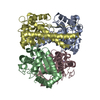
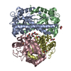
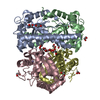
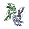
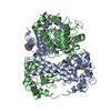
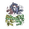
| Title | Crystal structure of superoxide dismutase from Pyrobaculum aerophilum | ||||||
 Components Components | Superoxide dismutase | ||||||
 Keywords Keywords | OXIDOREDUCTASE / ALPHA-BETA | ||||||
| Function / homology |  Function and homology information Function and homology informationsuperoxide dismutase / superoxide dismutase activity / metal ion binding Similarity search - Function | ||||||
| Biological species |   Pyrobaculum aerophilum (archaea) Pyrobaculum aerophilum (archaea) | ||||||
| Method |  X-RAY DIFFRACTION / SYNCHROTRON / MOLECULAR REPLACEMENT / Resolution: 1.8 Å X-RAY DIFFRACTION / SYNCHROTRON / MOLECULAR REPLACEMENT / Resolution: 1.8 Å | ||||||
 Authors Authors | Lee, S. / Sawaya, M.R. / Eisenberg, D. | ||||||
 Citation Citation | Journal: Acta Crystallogr.,Sect.D / Year: 2003 Title: Structure of superoxide dismutase from Pyrobaculum aerophilum presents a challenging case in molecular replacement with multiple molecules, pseudo-symmetry and twinning. Authors: Lee, S. / Sawaya, M.R. / Eisenberg, D. | ||||||
| History |
|
- Structure visualization
| Structure viewer | Molecule: MolmilJmol/JSmol |
|---|
- Downloads & links
-Download
| PDBx/mmCIF format | 1p7g.cif.gz | 1 MB | Display | PDBx/mmCIF format |
|---|---|---|---|---|
| PDB format | pdb1p7g.ent.gz | 868.2 KB | Display | PDB format |
| PDBx/mmJSON format | 1p7g.json.gz | Tree view | PDBx/mmJSON format | |
| Others |  Other downloads Other downloads |
-Validation report
| Arichive directory | https://data.pdbj.org/pub/pdb/validation_reports/p7/1p7gftp://data.pdbj.org/pub/pdb/validation_reports/p7/1p7g | HTTPS FTP |
|---|
-Related structure data
| Related structure data |  1sss 1iks S: Starting model for refinement |
|---|---|
| Similar structure data |


-Links
 PDBj
PDBj


- Assembly
| Deposited unit | 
| ||||||||
|---|---|---|---|---|---|---|---|---|---|
| 1 | 
| ||||||||
| 2 | 
| ||||||||
| 3 | 
| ||||||||
| 4 | 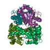
| ||||||||
| 5 | 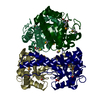
| ||||||||
| 6 | 
| ||||||||
| Unit cell |
| ||||||||
| Details | The biological assembly is a tetramer. There are six tetramers in the asymmetric unit. |
-Components
| #1: Protein | Mass: 25651.740 Da / Num. of mol.: 24 Source method: isolated from a genetically manipulated source Source: (gene. exp.) Pyrobaculum aerophilum (archaea) / Gene: SOD OR PAE0274 / Plasmid: pQE-30 / Species (production host): Escherichia coli / Production host:  #2: Chemical | ChemComp-ACT /   Mass: 59.044 Da / Num. of mol.: 19 / Source method: obtained synthetically / Formula: C2H3O2 Mass: 59.044 Da / Num. of mol.: 19 / Source method: obtained synthetically / Formula: C2H3O2#3: Chemical | ChemComp-BME /   Mass: 78.133 Da / Num. of mol.: 12 / Source method: obtained synthetically / Formula: C2H6OS Mass: 78.133 Da / Num. of mol.: 12 / Source method: obtained synthetically / Formula: C2H6OS#4: Water | ChemComp-HOH / |  Mass: 18.015 Da / Num. of mol.: 2536 / Source method: isolated from a natural source / Formula: H2O Mass: 18.015 Da / Num. of mol.: 2536 / Source method: isolated from a natural source / Formula: H2O |
|---|
-Experimental details
-Experiment
| Experiment | Method: X-RAY DIFFRACTION / Number of used crystals: 1 |
|---|
- Sample preparation
| Crystal | Density Matthews: 2.14 Å3/Da / Density % sol: 42.8 % | ||||||||||||||||||||||||||||||
|---|---|---|---|---|---|---|---|---|---|---|---|---|---|---|---|---|---|---|---|---|---|---|---|---|---|---|---|---|---|---|---|
| Crystal grow | Temperature: 298 K / Method: vapor diffusion, hanging drop / pH: 7.5 Details: PEG 3000, calcium acetate, hepes, pH 7.5, VAPOR DIFFUSION, HANGING DROP, temperature 298K | ||||||||||||||||||||||||||||||
| Crystal grow | *PLUS Temperature: 295 K / Method: vapor diffusion, hanging drop | ||||||||||||||||||||||||||||||
| Components of the solutions | *PLUS
|
-Data collection
| Diffraction | Mean temperature: 100 K | |||||||||||||||
|---|---|---|---|---|---|---|---|---|---|---|---|---|---|---|---|---|
| Diffraction source | Source: SYNCHROTRON / Site: NSLS  / Beamline: X8C / Wavelength: 0.9785, 0.9708, 0.9787, 0.9863 / Beamline: X8C / Wavelength: 0.9785, 0.9708, 0.9787, 0.9863 | |||||||||||||||
| Detector | Type: ADSC QUANTUM 4 / Detector: CCD / Date: Jun 11, 1999 / Details: MIRRORS | |||||||||||||||
| Radiation | Monochromator: MIRRORS / Protocol: MAD / Monochromatic (M) / Laue (L): M / Scattering type: x-ray | |||||||||||||||
| Radiation wavelength |
| |||||||||||||||
| Reflection | Resolution: 1.8→36.51 Å / Num. all: 476704 / Num. obs: 465126 / % possible obs: 97.6 % / Observed criterion σ(F): 0 / Observed criterion σ(I): 0 / Redundancy: 4.3 % / Biso Wilson estimate: 18.2 Å2 / Rmerge(I) obs: 0.076 / Net I/σ(I): 20.1 | |||||||||||||||
| Reflection shell | Resolution: 1.8→1.86 Å / Redundancy: 1.5 % / Rmerge(I) obs: 0.302 / Mean I/σ(I) obs: 3.3 / Num. unique all: 39384 / % possible all: 82.6 | |||||||||||||||
| Reflection | *PLUS Highest resolution: 1.8 Å / Lowest resolution: 36.4 Å / Num. measured all: 2013670 | |||||||||||||||
| Reflection shell | *PLUS % possible obs: 82.6 % |
- Processing
| Software |
| |||||||||||||||||||||||||
|---|---|---|---|---|---|---|---|---|---|---|---|---|---|---|---|---|---|---|---|---|---|---|---|---|---|---|
| Refinement | Method to determine structure: MOLECULAR REPLACEMENT Starting model: PDB ENTRY 1SSS 1sss Resolution: 1.8→36.45 Å / Isotropic thermal model: RESTRAINED / Cross valid method: THROUGHOUT / σ(F): 0 / Stereochemistry target values: Engh & Huber Details: Partial twinning incorporated with the twinning fraction being 0.463.
| |||||||||||||||||||||||||
| Displacement parameters | Biso mean: 21 Å2
| |||||||||||||||||||||||||
| Refine analyze |
| |||||||||||||||||||||||||
| Refinement step | Cycle: LAST / Resolution: 1.8→36.45 Å
| |||||||||||||||||||||||||
| Refine LS restraints |
| |||||||||||||||||||||||||
| LS refinement shell | Resolution: 1.8→1.86 Å
| |||||||||||||||||||||||||
| Refinement | *PLUS Highest resolution: 1.8 Å | |||||||||||||||||||||||||
| Solvent computation | *PLUS | |||||||||||||||||||||||||
| Displacement parameters | *PLUS | |||||||||||||||||||||||||
| Refine LS restraints | *PLUS
| |||||||||||||||||||||||||
| LS refinement shell | *PLUS Rfactor Rfree: 0.263 / Rfactor Rwork: 0.241 |
