Resolution: 1.85→41.54 Å / Cor.coef. Fo:Fc: 0.954 / Cor.coef. Fo:Fc free: 0.941 / SU B: 7.201 / SU ML: 0.107 / TLS residual ADP flag: LIKELY RESIDUAL / Cross valid method: THROUGHOUT / σ(F): 0 / ESU R: 0.166 / ESU R Free: 0.145 / Stereochemistry target values: MAXIMUM LIKELIHOOD Details: HYDROGENS HAVE BEEN ADDED IN THE RIDING POSITIONS; the region around GLU D 37 is highly flexible and poorly defined
Rfactor
Num. reflection
% reflection
Selection details
Rfree
0.23027
7500
10.1 %
RANDOM
Rwork
0.20159
-
-
-
all
0.20446
67009
-
-
obs
0.20446
67009
96.62 %
-
Solvent computation
Ion probe radii: 0.8 Å / Shrinkage radii: 0.8 Å / VDW probe radii: 1.2 Å / Solvent model: MASK
Displacement parameters
Biso mean: 30.565 Å2
Baniso -1
Baniso -2
Baniso -3
1-
0.06 Å2
0.03 Å2
0 Å2
2-
-
0.06 Å2
0 Å2
3-
-
-
-0.09 Å2
Refinement step
Cycle: LAST / Resolution: 1.85→41.54 Å
Protein
Nucleic acid
Ligand
Solvent
Total
Num. atoms
6856
0
4
177
7037
Refine LS restraints
Refine-ID
Type
Dev ideal
Dev ideal target
Number
X-RAY DIFFRACTION
r_bond_refined_d
0.009
0.022
7048
X-RAY DIFFRACTION
r_angle_refined_deg
1.08
1.938
9556
X-RAY DIFFRACTION
r_dihedral_angle_1_deg
6.027
5
840
X-RAY DIFFRACTION
r_dihedral_angle_2_deg
36.316
24.607
356
X-RAY DIFFRACTION
r_dihedral_angle_3_deg
13.062
15
1192
X-RAY DIFFRACTION
r_dihedral_angle_4_deg
13.451
15
28
X-RAY DIFFRACTION
r_chiral_restr
0.079
0.2
992
X-RAY DIFFRACTION
r_gen_planes_refined
0.004
0.02
5452
X-RAY DIFFRACTION
r_nbd_refined
0.188
0.2
3402
X-RAY DIFFRACTION
r_nbtor_refined
0.299
0.2
4821
X-RAY DIFFRACTION
r_xyhbond_nbd_refined
0.22
0.2
324
X-RAY DIFFRACTION
r_symmetry_vdw_refined
0.198
0.2
33
X-RAY DIFFRACTION
r_symmetry_hbond_refined
0.129
0.2
8
X-RAY DIFFRACTION
r_mcbond_it
0.473
1.5
4342
X-RAY DIFFRACTION
r_mcangle_it
0.729
2
6704
X-RAY DIFFRACTION
r_scbond_it
1.173
3
3177
X-RAY DIFFRACTION
r_scangle_it
1.748
4.5
2852
LS refinement shell
Resolution: 1.85→1.898 Å / Total num. of bins used: 20
Rfactor
Num. reflection
% reflection
Rfree
0.307
506
-
Rwork
0.255
4654
-
obs
-
-
91.72 %
Refinement TLS params.
Method: refined / Refine-ID: X-RAY DIFFRACTION
ID
L11 (°2)
L12 (°2)
L13 (°2)
L22 (°2)
L23 (°2)
L33 (°2)
S11 (Å °)
S12 (Å °)
S13 (Å °)
S21 (Å °)
S22 (Å °)
S23 (Å °)
S31 (Å °)
S32 (Å °)
S33 (Å °)
T11 (Å2)
T12 (Å2)
T13 (Å2)
T22 (Å2)
T23 (Å2)
T33 (Å2)
Origin x (Å)
Origin y (Å)
Origin z (Å)
1
2.0419
0.038
-0.2026
2.2394
0.7829
2.2565
-0.0037
0.2017
-0.2261
-0.1542
0.1978
-0.079
0.1491
0.0726
-0.1941
-0.1536
-0.0147
-0.0102
-0.189
-0.0509
-0.1897
100.7018
68.1653
-21.5696
2
1.8843
0.4302
0.7818
2.6596
1.8628
3.4263
-0.1013
0.183
-0.0538
-0.0666
0.6622
-0.8198
-0.2526
0.8473
-0.5609
-0.1607
-0.0663
0.0274
0.0405
-0.273
0.1014
123.8467
93.3193
-4.0409
3
2.0104
0.5548
0.9273
2.6384
0.9619
2.411
-0.1211
-0.1749
0.2001
-0.0763
0.0964
0.0889
-0.3251
-0.0715
0.0247
-0.1471
0.0357
-0.0093
-0.2022
-0.0302
-0.2082
100.2236
94.6506
-6.8439
4
1.8539
0.2316
0.1225
2.8473
2.0356
3.8714
-0.0064
0.1747
-0.2514
-0.1927
0.794
-1.0265
0.0485
1.1137
-0.7877
-0.1584
0.0194
0.058
0.1855
-0.4105
0.2574
124.443
69.3008
-22.4855
Refinement TLS group
ID
Refine-ID
Refine TLS-ID
Auth asym-ID
Auth seq-ID
1
X-RAY DIFFRACTION
1
A
12 - 222
2
X-RAY DIFFRACTION
2
B
12 - 222
3
X-RAY DIFFRACTION
3
C
12 - 222
4
X-RAY DIFFRACTION
4
D
12 - 222
+
About Yorodumi
-
News
-
Feb 9, 2022. New format data for meta-information of EMDB entries
New format data for meta-information of EMDB entries
Version 3 of the EMDB header file is now the official format.
The previous official version 1.9 will be removed from the archive.
In the structure databanks used in Yorodumi, some data are registered as the other names, "COVID-19 virus" and "2019-nCoV". Here are the details of the virus and the list of structure data.
Jan 31, 2019. EMDB accession codes are about to change! (news from PDBe EMDB page)
EMDB accession codes are about to change! (news from PDBe EMDB page)
The allocation of 4 digits for EMDB accession codes will soon come to an end. Whilst these codes will remain in use, new EMDB accession codes will include an additional digit and will expand incrementally as the available range of codes is exhausted. The current 4-digit format prefixed with “EMD-” (i.e. EMD-XXXX) will advance to a 5-digit format (i.e. EMD-XXXXX), and so on. It is currently estimated that the 4-digit codes will be depleted around Spring 2019, at which point the 5-digit format will come into force.
The EM Navigator/Yorodumi systems omit the EMD- prefix.
Related info.:Q: What is EMD? / ID/Accession-code notation in Yorodumi/EM Navigator
Yorodumi is a browser for structure data from EMDB, PDB, SASBDB, etc.
This page is also the successor to EM Navigator detail page, and also detail information page/front-end page for Omokage search.
The word "yorodu" (or yorozu) is an old Japanese word meaning "ten thousand". "mi" (miru) is to see.
Related info.:EMDB / PDB / SASBDB / Comparison of 3 databanks / Yorodumi Search / Aug 31, 2016. New EM Navigator & Yorodumi / Yorodumi Papers / Jmol/JSmol / Function and homology information / Changes in new EM Navigator and Yorodumi
 Movie
Movie Controller

 Yorodumi
 Open data
 Basic information
 Components
 Keywords
 Function and homology information
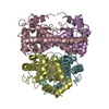
 Pyrobaculum aerophilum (archaea)
 X-RAY DIFFRACTION /
  Authors
 Citation
 Structure visualization
 Downloads & links
 Other downloads


 PDBj
PDBj
 Assembly
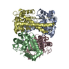


 Mass: 54.938 Da / Num. of mol.: 4 / Source method: obtained synthetically / Formula: Mn
 Mass: 18.015 Da / Num. of mol.: 177 / Source method: isolated from a natural source / Formula: H2O
 Sample preparation
 / Beamline: X8C / Wavelength: 1.1 Å
 Processing