 Movie
Movie Controller

[English] 日本語
 Yorodumi
- PDB-1gn3: H145Q mutant of Mycobacterium tuberculosis iron-superoxide dismutase. -
+ Open data

- Basic information
| Entry | Database: PDB / ID: 1gn3 | ||||||
|---|---|---|---|---|---|---|---|
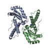
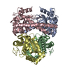
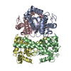
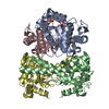
| Title | H145Q mutant of Mycobacterium tuberculosis iron-superoxide dismutase. | ||||||
 Components Components | SUPEROXIDE DISMUTASE | ||||||
 Keywords Keywords | OXIDOREDUCTASE | ||||||
| Function / homology |  Function and homology information Function and homology informationTolerance of reactive oxygen produced by macrophages / detoxification / superoxide dismutase / superoxide dismutase activity / manganese ion binding / response to oxidative stress / periplasmic space / iron ion binding / extracellular region / plasma membrane / cytosol Similarity search - Function | ||||||
| Biological species |   MYCOBACTERIUM TUBERCULOSIS (bacteria) MYCOBACTERIUM TUBERCULOSIS (bacteria) | ||||||
| Method |  X-RAY DIFFRACTION / MOLECULAR REPLACEMENT / Resolution: 4 Å X-RAY DIFFRACTION / MOLECULAR REPLACEMENT / Resolution: 4 Å | ||||||
 Authors Authors | Bunting, K.A. / Cooper, J.B. / Badasso, M.O. / Tickle, I.J. / Newton, M. / Wood, S.P. / Zhang, Y. / Young, D.B. | ||||||
 Citation Citation | Journal: Eur.J.Biochem. / Year: 1998 Title: Engineering a Change in the Metal-Ion Specificity of the Iron-Depedent Superoxide Dismutase from Mycobacterium Tuberculosis. X-Ray Structure Analysis of Site-Directed Mutants. Authors: Bunting, K.A. / Cooper, J.B. / Badasso, M.O. / Tickle, I.J. / Newton, M. / Wood, S.P. / Young, D.B. | ||||||
| History |
|
- Structure visualization
| Structure viewer | Molecule: MolmilJmol/JSmol |
|---|
- Downloads & links
-Download
| PDBx/mmCIF format | 1gn3.cif.gz | 86.6 KB | Display | PDBx/mmCIF format |
|---|---|---|---|---|
| PDB format | pdb1gn3.ent.gz | 65.2 KB | Display | PDB format |
| PDBx/mmJSON format | 1gn3.json.gz | Tree view | PDBx/mmJSON format | |
| Others |  Other downloads Other downloads |
-Validation report
| Arichive directory | https://data.pdbj.org/pub/pdb/validation_reports/gn/1gn3ftp://data.pdbj.org/pub/pdb/validation_reports/gn/1gn3 | HTTPS FTP |
|---|
-Related structure data
| Related structure data |  1gn4C  1idsS C: citing same article ( S: Starting model for refinement |
|---|---|
| Similar structure data |


-Links
 PDBj
PDBj
- Assembly
| Deposited unit | 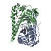
| ||||||||
|---|---|---|---|---|---|---|---|---|---|
| 1 | 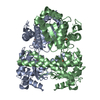
| ||||||||
| Unit cell |
| ||||||||
| Noncrystallographic symmetry (NCS) | NCS oper: (Code: given Matrix: (-0.9936, -0.0017, 0.1129), Vector: |
-Components
| #1: Protein | Mass: 23050.844 Da / Num. of mol.: 2 / Mutation: YES Source method: isolated from a genetically manipulated source Source: (gene. exp.) MYCOBACTERIUM TUBERCULOSIS (bacteria) / Production host: MYCOBACTERIUM VACCAE (bacteria)References: UniProt: P17670, UniProt: P9WGE7*PLUS, superoxide dismutase #2: Chemical |   Mass: 55.845 Da / Num. of mol.: 2 / Source method: obtained synthetically / Formula: Fe Mass: 55.845 Da / Num. of mol.: 2 / Source method: obtained synthetically / Formula: Fe |
|---|
-Experimental details
-Experiment
| Experiment | Method: X-RAY DIFFRACTION / Number of used crystals: 1 |
|---|
- Sample preparation
| Crystal | Density Matthews: 2.36 Å3/Da / Density % sol: 48 % |
|---|---|
| Crystal grow | Temperature: 277 K / pH: 7 Details: 100MM TRIS-HCL PH 7.0, 25% PEG 6000, PROTEIN CONCENTRATION = 3 MG/ML, 4 DEGREES CENTIGRADE. |
-Data collection
| Diffraction | Mean temperature: 293 K |
|---|---|
| Diffraction source | Source: ROTATING ANODE / Type: ENRAF-NONIUS FR591 / Wavelength: 1.5418 |
| Detector | Type: MARRESEARCH / Detector: IMAGE PLATE |
| Radiation | Monochromator: GRAPHITE / Protocol: SINGLE WAVELENGTH / Monochromatic (M) / Laue (L): M / Scattering type: x-ray |
| Radiation wavelength | Wavelength: 1.5418 Å / Relative weight: 1 |
| Reflection | Resolution: 4→99 Å / Num. obs: 5069 / % possible obs: 98.4 % / Redundancy: 9.2 % / Rmerge(I) obs: 0.163 |
| Reflection shell | Rmerge(I) obs: 0.221 |
- Processing
| Software |
| |||||||||||||||||||||||||||||||||||||||||||||||||||||||||||||||
|---|---|---|---|---|---|---|---|---|---|---|---|---|---|---|---|---|---|---|---|---|---|---|---|---|---|---|---|---|---|---|---|---|---|---|---|---|---|---|---|---|---|---|---|---|---|---|---|---|---|---|---|---|---|---|---|---|---|---|---|---|---|---|---|---|
| Refinement | Method to determine structure: MOLECULAR REPLACEMENT Starting model: PDB ENTRY 1IDS Resolution: 4→20 Å / Cross valid method: THROUGHOUT / σ(F): 0
| |||||||||||||||||||||||||||||||||||||||||||||||||||||||||||||||
| Refinement step | Cycle: LAST / Resolution: 4→20 Å
| |||||||||||||||||||||||||||||||||||||||||||||||||||||||||||||||
| Refine LS restraints |
|
