 Movie
Movie Controller


+ Open data

- Basic information
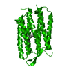
| Entry | Database: PDB / ID: 1brx | |||||||||
|---|---|---|---|---|---|---|---|---|---|---|
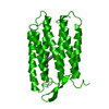
| Title | BACTERIORHODOPSIN/LIPID COMPLEX | |||||||||
 Components Components | BACTERIORHODOPSIN | |||||||||
 Keywords Keywords | PROTON PUMP / MEMBRANE PROTEIN / RETINAL PROTEIN / LIPIDS / PHOTORECEPTOR / HALOARCHAEA | |||||||||
| Function / homology |  Function and homology informationphotoreceptor activity / phototransduction / proton transmembrane transport / monoatomic ion channel activity / plasma membrane Function and homology informationphotoreceptor activity / phototransduction / proton transmembrane transport / monoatomic ion channel activity / plasma membraneSimilarity search - Function | |||||||||
| Biological species |  Halobacterium salinarum (Halophile) Halobacterium salinarum (Halophile) | |||||||||
| Method | X-RAY DIFFRACTION / MOLECULAR REPLACEMENT / Resolution: 2.3 Å | |||||||||
 Authors Authors | Luecke, H. / Richter, H.T. / Lanyi, J. | |||||||||
 Citation Citation | Journal: Science / Year: 1998 Title: Proton transfer pathways in bacteriorhodopsin at 2.3 angstrom resolution. Authors: Luecke, H. / Richter, H.T. / Lanyi, J.K. | |||||||||
| History |
|
- Structure visualization
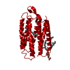
| Structure viewer | Molecule: MolmilJmol/JSmol |
|---|
- Downloads & links
-Download
| PDBx/mmCIF format | 1brx.cif.gz | 48.8 KB | Display | PDBx/mmCIF format |
|---|---|---|---|---|
| PDB format | pdb1brx.ent.gz | 37.6 KB | Display | PDB format |
| PDBx/mmJSON format | 1brx.json.gz | Tree view | PDBx/mmJSON format | |
| Others |  Other downloads Other downloads |
-Validation report
| Arichive directory | https://data.pdbj.org/pub/pdb/validation_reports/br/1brxftp://data.pdbj.org/pub/pdb/validation_reports/br/1brx | HTTPS FTP |
|---|
-Related structure data


-Links
 PDBj
PDBj


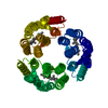
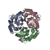
- Assembly
| Deposited unit | 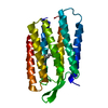
| ||||||||
|---|---|---|---|---|---|---|---|---|---|
| 1 | 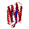
| ||||||||
| Unit cell |
|
-Components
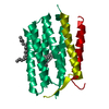
| #1: Protein | Mass: 26797.381 Da / Num. of mol.: 1 Source method: isolated from a genetically manipulated source Details: SCHIFF BASE LINKAGE BETWEEN LYS 126 (NZ) AND RET 301 (C15) Source: (gene. exp.) Halobacterium salinarum (Halophile) / Cellular location: PLASMA MEMBRANECell membrane / Organ: PLASMA / Cellular location (production host): CYTOPLASM / Production host: Halobacterium salinarum (Halophile) / References: UniProt: P02945 |
|---|---|
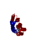
| #2: Chemical | ChemComp-RET / Retinal  Mass: 284.436 Da / Num. of mol.: 1 / Source method: obtained synthetically / Formula: C20H28O Mass: 284.436 Da / Num. of mol.: 1 / Source method: obtained synthetically / Formula: C20H28O |
| #3: Water | ChemComp-HOH / Water Mass: 18.015 Da / Num. of mol.: 3 / Source method: isolated from a natural source / Formula: H2O Mass: 18.015 Da / Num. of mol.: 3 / Source method: isolated from a natural source / Formula: H2O |
-Experimental details
-Experiment
| Experiment | Method: X-RAY DIFFRACTION / Number of used crystals: 1 |
|---|
- Sample preparation
| Crystal | Density Matthews: 2.23 Å3/Da / Density % sol: 43.08 % / Description: FLASH-COOLED IN LN2 STREAM | ||||||||||||||||||||
|---|---|---|---|---|---|---|---|---|---|---|---|---|---|---|---|---|---|---|---|---|---|
| Crystal grow | pH: 5.6 / Details: pH 5.6 | ||||||||||||||||||||
| Crystal grow | *PLUS Temperature: 20 ℃ / Method: unknown | ||||||||||||||||||||
| Components of the solutions | *PLUS
|
-Data collection
| Diffraction | Mean temperature: 100 K |
|---|---|
| Diffraction source | Source: ROTATING ANODE / Type: RIGAKU RUH3R / Wavelength: 1.5418 |
| Detector | Type: RIGAKU RAXIS IV / Detector: IMAGE PLATE / Date: Oct 1, 1997 / Details: MIRRORS |
| Radiation | Monochromator: MIRROR / Monochromatic (M) / Laue (L): M / Scattering type: x-ray |
| Radiation wavelength | Wavelength: 1.5418 Å / Relative weight: 1 |
| Reflection | Resolution: 2.3→25 Å / Num. obs: 9769 / % possible obs: 96.5 % / Observed criterion σ(I): 0 / Redundancy: 25.6 % / Rsym value: 0.113 / Net I/σ(I): 15.1 |
| Reflection shell | Resolution: 2.3→2.34 Å / Mean I/σ(I) obs: 1.7 / Rsym value: 0.443 / % possible all: 75.9 |
| Reflection | *PLUS Num. measured all: 250474 / Rmerge(I) obs: 0.113 |
| Reflection shell | *PLUS % possible obs: 75.9 % / Rmerge(I) obs: 0.443 |
- Processing
| Software |
| |||||||||||||||||||||||||||||||||
|---|---|---|---|---|---|---|---|---|---|---|---|---|---|---|---|---|---|---|---|---|---|---|---|---|---|---|---|---|---|---|---|---|---|---|
| Refinement | Method to determine structure: MOLECULAR REPLACEMENT Starting model: 1AT9 AND 2BRD Resolution: 2.3→12 Å / Num. parameters: 6544 / Num. restraintsaints: 6912 / Cross valid method: FREE R-VALUE / σ(F): 0 / Stereochemistry target values: ENGH AND HUBER / Details: USED TWIN OPTION, REFINED TWIN RATIO: 0.54/0.46.
| |||||||||||||||||||||||||||||||||
| Solvent computation | Solvent model: SWAT | |||||||||||||||||||||||||||||||||
| Refine analyze | Num. disordered residues: 0 | |||||||||||||||||||||||||||||||||
| Refinement step | Cycle: LAST / Resolution: 2.3→12 Å
| |||||||||||||||||||||||||||||||||
| Refine LS restraints |
| |||||||||||||||||||||||||||||||||
| Software | *PLUS Name: SHELXL-97 / Classification: refinement | |||||||||||||||||||||||||||||||||
| Refinement | *PLUS σ(F): 4 / Rfactor all: 0.223 / Rfactor obs: 0.18 | |||||||||||||||||||||||||||||||||
| Solvent computation | *PLUS | |||||||||||||||||||||||||||||||||
| Displacement parameters | *PLUS |
