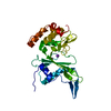
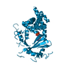
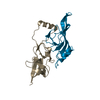
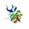
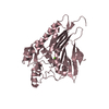
Entry Database : PDB / ID : 1bdyTitle C2 DOMAIN FROM PROTEIN KINASE C DELTA PROTEIN KINASE C Keywords / / / / / / Function / homology Function Domain/homology Component
/ / / / / / / / / / / / / / / / / / / / / / / / / / / / / / / / / / / / / / / / / / / / / / / / / / / / / / / / / / / / / / / / / / / / / / / / / / / / / / / / / / / / / / / / / / / / / / / / / / / / / / / / / / / / / / / / / / / / / / / / / / / / / / / / / / / / / / / / / / Biological species Rattus norvegicus (Norway rat)Method / / / Resolution : 2.2 Å Authors Pappa, H. / Murray-Rust, J. / Dekker, L.V. / Parker, P.J. / Mcdonald, N.Q. Journal : Structure / Year : 1998Title : Crystal structure of the C2 domain from protein kinase C-delta.Authors : Pappa, H. / Murray-Rust, J. / Dekker, L.V. / Parker, P.J. / McDonald, N.Q. History Deposition May 11, 1998 Processing site Revision 1.0 Oct 14, 1998 Provider / Type Revision 1.1 Mar 24, 2008 Group Revision 1.2 Jul 13, 2011 Group Revision 1.3 Feb 7, 2024 Group / Database references / OtherCategory chem_comp_atom / chem_comp_bond ... chem_comp_atom / chem_comp_bond / database_2 / pdbx_database_status Item / _database_2.pdbx_database_accession / _pdbx_database_status.process_site
Show all Show less
 Movie
Movie Controller


 Open data
 Basic information
 Components
 Keywords
 Function and homology information
 X-RAY DIFFRACTION /
  Authors
 Citation
 Structure visualization
 Downloads & links
 Other downloads
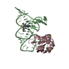
 PDBj
PDBj


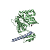
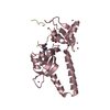
 Assembly
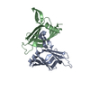

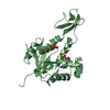
 Mass: 18.015 Da / Num. of mol.: 82 / Source method: isolated from a natural source / Formula: H2O
 Sample preparation
 / Beamline: PX9.6 / Wavelength: 0.87
  Processing