 Movie
Movie Controller


+ Open data

- Basic information
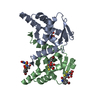
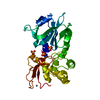
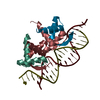
| Entry | Database: PDB / ID: 1b01 | ||||||
|---|---|---|---|---|---|---|---|
| Title | TRANSCRIPTIONAL REPRESSOR COPG/DNA COMPLEX | ||||||
 Components Components |
| ||||||
 Keywords Keywords | GENE REGULATION/DNA / TRANSCRIPTIONAL REPRESSOR / DNA-BINDING PROTEIN / PLASMID / PROTEIN-DNA COMPLEX / GENE REGULATION-DNA complex | ||||||
| Function / homology |  Function and homology information Function and homology informationplasmid maintenance / protein-DNA complex / sequence-specific DNA binding / regulation of DNA-templated transcription Similarity search - Function | ||||||
| Biological species |  Streptococcus agalactiae (bacteria) Streptococcus agalactiae (bacteria) | ||||||
| Method |  X-RAY DIFFRACTION / SYNCHROTRON / MIRAS / Resolution: 2.56 Å X-RAY DIFFRACTION / SYNCHROTRON / MIRAS / Resolution: 2.56 Å | ||||||
 Authors Authors | Gomis-Rueth, F.X. / Sola, M. / Acebo, P. / Parraga, A. / Guasch, A. / Eritja, R. / Gonzalez, A. / Espinosa, M. / del Solar, G. / Coll, M. | ||||||
 Citation Citation | Journal: EMBO J. / Year: 1998 Title: The structure of plasmid-encoded transcriptional repressor CopG unliganded and bound to its operator. Authors: Gomis-Ruth, F.X. / Sola, M. / Acebo, P. / Parraga, A. / Guasch, A. / Eritja, R. / Gonzalez, A. / Espinosa, M. / del Solar, G. / Coll, M. #1: Journal: FEBS Lett. / Year: 1998Title: Overexpression, Purification, Crystallization and Preliminary X-Ray Diffraction Analysis of the Pmv158-Encoded Plasmid Transcriptional Repressor Protein Copg Authors: Gomis-Rueth, F.X. / Sola, M. / Perez-Luque, R. / Acebo, P. / Alda, M.T. / Gonzalez, A. / Espinosa, M. / del Solar, G. / Coll, M. | ||||||
| History |
|
- Structure visualization
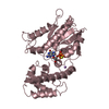
| Structure viewer | Molecule: MolmilJmol/JSmol |
|---|
- Downloads & links
-Download
| PDBx/mmCIF format | 1b01.cif.gz | 49.2 KB | Display | PDBx/mmCIF format |
|---|---|---|---|---|
| PDB format | pdb1b01.ent.gz | 34.4 KB | Display | PDB format |
| PDBx/mmJSON format | 1b01.json.gz | Tree view | PDBx/mmJSON format | |
| Others |  Other downloads Other downloads |
-Validation report
| Arichive directory | https://data.pdbj.org/pub/pdb/validation_reports/b0/1b01ftp://data.pdbj.org/pub/pdb/validation_reports/b0/1b01 | HTTPS FTP |
|---|
-Related structure data


-Links
 PDBj
PDBj


- Assembly
| Deposited unit | 
| ||||||||||
|---|---|---|---|---|---|---|---|---|---|---|---|
| 1 | 
| ||||||||||
| Unit cell |
| ||||||||||
| Noncrystallographic symmetry (NCS) | NCS oper: (Code: given Matrix: (-0.36851, -0.861028, -0.350473), Vector: |
-Components
| #1: DNA chain | Mass: 5749.738 Da / Num. of mol.: 1 / Source method: obtained synthetically | ||
|---|---|---|---|
| #2: DNA chain | Mass: 5900.820 Da / Num. of mol.: 1 / Source method: obtained synthetically | ||
| #3: Protein/peptide | Mass: 5124.092 Da / Num. of mol.: 2 / Fragment: DNA-BINDING PROTEIN Source method: isolated from a genetically manipulated source Source: (gene. exp.) Streptococcus agalactiae (bacteria) / Strain: PLS1 / Cellular location: PLASMID PMV158 / Plasmid: PMV158 / Production host: #4: Water | ChemComp-HOH / |  Mass: 18.015 Da / Num. of mol.: 13 / Source method: isolated from a natural source / Formula: H2O Mass: 18.015 Da / Num. of mol.: 13 / Source method: isolated from a natural source / Formula: H2O |
-Experimental details
-Experiment
| Experiment | Method: X-RAY DIFFRACTION / Number of used crystals: 3 |
|---|
- Sample preparation
| Crystal | Density Matthews: 2.23 Å3/Da / Density % sol: 45 % | ||||||||||||||||||||
|---|---|---|---|---|---|---|---|---|---|---|---|---|---|---|---|---|---|---|---|---|---|
| Crystal grow | Method: vapor diffusion, hanging drop / pH: 6.7 / Details: MPD, HEPES, pH 6.70, VAPOR DIFFUSION, HANGING DROP | ||||||||||||||||||||
| Components of the solutions |
| ||||||||||||||||||||
| Crystal | *PLUS | ||||||||||||||||||||
| Crystal grow | *PLUS PH range low: 7.5 / PH range high: 6.7 | ||||||||||||||||||||
| Components of the solutions | *PLUS
|
-Data collection
| Diffraction | Mean temperature: 100 K |
|---|---|
| Diffraction source | Source: SYNCHROTRON / Site: EMBL/DESY, HAMBURG  / Beamline: X31 / Wavelength: 1.5418 / Beamline: X31 / Wavelength: 1.5418 |
| Detector | Type: MARRESEARCH / Detector: IMAGE PLATE |
| Radiation | Monochromator: GRAPHITE / Protocol: SINGLE WAVELENGTH / Monochromatic (M) / Laue (L): M / Scattering type: x-ray |
| Radiation wavelength | Wavelength: 1.5418 Å / Relative weight: 1 |
| Reflection | Resolution: 1.6→19.61 Å / Num. obs: 91693 / % possible obs: 98.1 % / Redundancy: 5 % / Rmerge(I) obs: 0.08 / Net I/σ(I): 10.3 |
| Reflection shell | Resolution: 1.6→1.69 Å / Rmerge(I) obs: 0.126 / Mean I/σ(I) obs: 3 / % possible all: 96.4 |
| Reflection | *PLUS Highest resolution: 2.56 Å / Lowest resolution: 19.86 Å / Num. obs: 6252 / % possible obs: 96.3 % / Redundancy: 5.9 % / Num. measured all: 78954 / Rmerge(I) obs: 0.062 |
| Reflection shell | *PLUS Highest resolution: 2.56 Å / Lowest resolution: 2.7 Å / % possible obs: 86.8 % / Rmerge(I) obs: 0.455 / Mean I/σ(I) obs: 1.7 |
- Processing
| Software |
| ||||||||||||||||||||||||||||||||||||||||||||||||||||||||||||
|---|---|---|---|---|---|---|---|---|---|---|---|---|---|---|---|---|---|---|---|---|---|---|---|---|---|---|---|---|---|---|---|---|---|---|---|---|---|---|---|---|---|---|---|---|---|---|---|---|---|---|---|---|---|---|---|---|---|---|---|---|---|
| Refinement | Method to determine structure: MIRAS / Resolution: 2.56→20 Å / Cross valid method: THROUGHOUT / σ(F): 2 Details: THE COMPLEX SET UP FOR CRYSTALLIZATION WAS MADE UP BY A COPG DIMER-OF-DIMERS AND A 19-BP DSDNA. AS THE DNA MOIETY IS PRESENT IN DUAL OCCUPANCY, A CRYSTALLOGRAPHIC DYAD IS CREATED. ...Details: THE COMPLEX SET UP FOR CRYSTALLIZATION WAS MADE UP BY A COPG DIMER-OF-DIMERS AND A 19-BP DSDNA. AS THE DNA MOIETY IS PRESENT IN DUAL OCCUPANCY, A CRYSTALLOGRAPHIC DYAD IS CREATED. ACCORDINGLY, THE CRYSTALLOGRAPHIC ASYMMETRIC UNIT COMPRISES HALF A PROTEIN/DNA COMPLEX, THAT IS, A COPG DIMER AND HALF DSDNA. THE DNA PART HAS BEEN MODELLED WITH THE TWO OBSERVED ORIENTATIONS, EACH WITH OCCUPANCY 0.5. IN THIS MODEL, DNA CHAIN E PAIRS CHAIN F AND CHAIN G DOES SO WITH CHAIN H.
| ||||||||||||||||||||||||||||||||||||||||||||||||||||||||||||
| Refinement step | Cycle: LAST / Resolution: 2.56→20 Å
| ||||||||||||||||||||||||||||||||||||||||||||||||||||||||||||
| Refine LS restraints |
|
