 Movie
Movie Controller


+ Open data

- Basic information
| Entry | Database: PDB / ID: 6nwd | ||||||
|---|---|---|---|---|---|---|---|
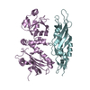
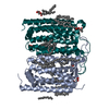
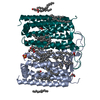
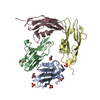
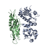
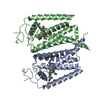
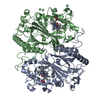
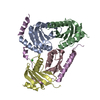
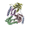
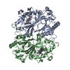
| Title | X-ray Crystallographic structure of Gloeobacter rhodopsin | ||||||
 Components Components | Gll0198 protein | ||||||
 Keywords Keywords | TRANSPORT PROTEIN / Proton pump / Photoreceptor | ||||||
| Function / homology |  Function and homology information Function and homology informationlight-activated monoatomic ion channel activity / photoreceptor activity / phototransduction / membrane / identical protein binding Similarity search - Function | ||||||
| Biological species |  Gloeobacter violaceus (bacteria) Gloeobacter violaceus (bacteria) | ||||||
| Method |  X-RAY DIFFRACTION / SYNCHROTRON / MOLECULAR REPLACEMENT / Resolution: 2 Å X-RAY DIFFRACTION / SYNCHROTRON / MOLECULAR REPLACEMENT / Resolution: 2 Å | ||||||
 Authors Authors | Ernst, O.P. / Morizumi, T. / Ou, W.L. | ||||||
| Funding support |  Canada, 1items Canada, 1items
| ||||||
 Citation Citation | Journal: Sci Rep / Year: 2019 Title: X-ray Crystallographic Structure and Oligomerization of Gloeobacter Rhodopsin. Authors: Morizumi, T. / Ou, W.L. / Van Eps, N. / Inoue, K. / Kandori, H. / Brown, L.S. / Ernst, O.P. | ||||||
| History |
|
- Structure visualization
| Structure viewer | Molecule: MolmilJmol/JSmol |
|---|
- Downloads & links
-Download
| PDBx/mmCIF format | 6nwd.cif.gz | 68.9 KB | Display | PDBx/mmCIF format |
|---|---|---|---|---|
| PDB format | pdb6nwd.ent.gz | 47.7 KB | Display | PDB format |
| PDBx/mmJSON format | 6nwd.json.gz | Tree view | PDBx/mmJSON format | |
| Others |  Other downloads Other downloads |
-Validation report
| Arichive directory | https://data.pdbj.org/pub/pdb/validation_reports/nw/6nwdftp://data.pdbj.org/pub/pdb/validation_reports/nw/6nwd | HTTPS FTP |
|---|
-Related structure data
| Related structure data |  3ddlS S: Starting model for refinement |
|---|---|
| Similar structure data |
-Links
 PDBj
PDBj


- Assembly
| Deposited unit | 
| ||||||||||||
|---|---|---|---|---|---|---|---|---|---|---|---|---|---|
| 1 | 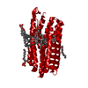
| ||||||||||||
| Unit cell |
| ||||||||||||
| Components on special symmetry positions |
|
-Components
-Protein , 1 types, 1 molecules A
| #1: Protein | Mass: 32307.518 Da / Num. of mol.: 1 Source method: isolated from a genetically manipulated source Source: (gene. exp.) Gloeobacter violaceus (strain ATCC 29082 / PCC 7421) (bacteria)Strain: ATCC 29082 / PCC 7421 / Gene: gll0198 / Production host: |
|---|
-Non-polymers , 5 types, 20 molecules 


| #2: Chemical | ChemComp-RET /  Mass: 284.436 Da / Num. of mol.: 1 / Source method: obtained synthetically / Formula: C20H28O / Feature type: SUBJECT OF INVESTIGATION Mass: 284.436 Da / Num. of mol.: 1 / Source method: obtained synthetically / Formula: C20H28O / Feature type: SUBJECT OF INVESTIGATION | ||||||
|---|---|---|---|---|---|---|---|
| #3: Chemical |  Mass: 678.940 Da / Num. of mol.: 3 / Source method: obtained synthetically / Formula: C36H73NO8P / Comment: DMPC, phospholipid*YM Mass: 678.940 Da / Num. of mol.: 3 / Source method: obtained synthetically / Formula: C36H73NO8P / Comment: DMPC, phospholipid*YM#4: Chemical | ChemComp-D10 / |  Mass: 142.282 Da / Num. of mol.: 1 / Source method: obtained synthetically / Formula: C10H22 Mass: 142.282 Da / Num. of mol.: 1 / Source method: obtained synthetically / Formula: C10H22#5: Chemical | ChemComp-D12 / |  Mass: 170.335 Da / Num. of mol.: 1 / Source method: obtained synthetically / Formula: C12H26 Mass: 170.335 Da / Num. of mol.: 1 / Source method: obtained synthetically / Formula: C12H26#6: Water | ChemComp-HOH / | Mass: 18.015 Da / Num. of mol.: 14 / Source method: isolated from a natural source / Formula: H2O |
-Details
| Has ligand of interest | Y |
|---|---|
| Has protein modification | Y |
-Experimental details
-Experiment
| Experiment | Method: X-RAY DIFFRACTION / Number of used crystals: 1 |
|---|
- Sample preparation
| Crystal | Density Matthews: 2.24 Å3/Da / Density % sol: 45.14 % |
|---|---|
| Crystal grow | Temperature: 307 K / Method: vapor diffusion, hanging drop / pH: 3.4 Details: 2.6-2.8 M NaH2PO4, 1,6-hexanediol, triethylene glycol, zinc acetate, n-Octyl-beta-D-Glucoside |
-Data collection
| Diffraction | Mean temperature: 100 K / Serial crystal experiment: N |
|---|---|
| Diffraction source | Source: SYNCHROTRON / Site: APS  / Beamline: 23-ID-D / Wavelength: 1.0332 Å / Beamline: 23-ID-D / Wavelength: 1.0332 Å |
| Detector | Type: DECTRIS PILATUS3 S 6M / Detector: PIXEL / Date: Mar 11, 2016 |
| Radiation | Protocol: SINGLE WAVELENGTH / Monochromatic (M) / Laue (L): M / Scattering type: x-ray |
| Radiation wavelength | Wavelength: 1.0332 Å / Relative weight: 1 |
| Reflection | Resolution: 2→42.79 Å / Num. obs: 19500 / % possible obs: 96.37 % / Redundancy: 1.8 % / CC1/2: 0.997 / Rmerge(I) obs: 0.04428 / Rpim(I) all: 0.04428 / Rrim(I) all: 0.06262 / Net I/σ(I): 6.6 |
| Reflection shell | Resolution: 2→2.072 Å / Rmerge(I) obs: 0.2155 / Num. unique obs: 19313 / CC1/2: 0.891 / Rpim(I) all: 0.2155 / Rrim(I) all: 0.3047 |
- Processing
| Software |
| ||||||||||||||||
|---|---|---|---|---|---|---|---|---|---|---|---|---|---|---|---|---|---|
| Refinement | Method to determine structure: MOLECULAR REPLACEMENT Starting model: 3DDL Resolution: 2→42.79 Å / Cross valid method: FREE R-VALUE
| ||||||||||||||||
| Refinement step | Cycle: LAST / Resolution: 2→42.79 Å
| ||||||||||||||||
| LS refinement shell | Resolution: 2→2.072 Å /
|
