 Movie
Movie Controller


+ Open data

- Basic information
| Entry | Database: PDB / ID: 6lty | ||||||
|---|---|---|---|---|---|---|---|
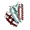
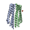
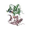
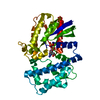
| Title | DNA bound antitoxin HigA3 | ||||||
 Components Components |
| ||||||
 Keywords Keywords | ANTITOXIN / DNA BINDING PROTEIN/DNA / DNA BINDING PROTEIN-DNA COMPLEX | ||||||
| Function / homology |  Function and homology information Function and homology information | ||||||
| Biological species |  Mycobacterium tuberculosis H37Rv (bacteria) Mycobacterium tuberculosis H37Rv (bacteria) | ||||||
| Method |  X-RAY DIFFRACTION / SYNCHROTRON / MOLECULAR REPLACEMENT / Resolution: 3.28 Å X-RAY DIFFRACTION / SYNCHROTRON / MOLECULAR REPLACEMENT / Resolution: 3.28 Å | ||||||
 Authors Authors | Park, J.Y. / Lee, B.J. | ||||||
| Funding support |  Korea, Republic Of, 1items Korea, Republic Of, 1items
| ||||||
 Citation Citation | Journal: Iucrj / Year: 2020 Title: Induced DNA bending by unique dimerization of HigA antitoxin. Authors: Park, J.Y. / Kim, H.J. / Pathak, C. / Yoon, H.J. / Kim, D.H. / Park, S.J. / Lee, B.J. | ||||||
| History |
|
- Structure visualization
| Structure viewer | Molecule: MolmilJmol/JSmol |
|---|
- Downloads & links
-Download
| PDBx/mmCIF format | 6lty.cif.gz | 67.3 KB | Display | PDBx/mmCIF format |
|---|---|---|---|---|
| PDB format | pdb6lty.ent.gz | 45.6 KB | Display | PDB format |
| PDBx/mmJSON format | 6lty.json.gz | Tree view | PDBx/mmJSON format | |
| Others |  Other downloads Other downloads |
-Validation report
| Arichive directory | https://data.pdbj.org/pub/pdb/validation_reports/lt/6ltyftp://data.pdbj.org/pub/pdb/validation_reports/lt/6lty | HTTPS FTP |
|---|
-Related structure data
| Related structure data |  6ltzSC S: Starting model for refinement C: citing same article ( |
|---|---|
| Similar structure data |


-Links
 PDBj
PDBj


- Assembly
| Deposited unit | 
| ||||||||
|---|---|---|---|---|---|---|---|---|---|
| 1 |
| ||||||||
| Unit cell |
|
-Components
| #1: Protein | Mass: 12891.620 Da / Num. of mol.: 2 Source method: isolated from a genetically manipulated source Source: (gene. exp.) Mycobacterium tuberculosis H37Rv (bacteria)Strain: H37Rv / Gene: higA3, Rv3183 / Production host: #2: DNA chain | | Mass: 6136.008 Da / Num. of mol.: 1 / Source method: obtained synthetically Source: (synth.) Mycobacterium tuberculosis H37Rv (bacteria)#3: DNA chain | | Mass: 6130.961 Da / Num. of mol.: 1 / Source method: obtained synthetically Source: (synth.) Mycobacterium tuberculosis H37Rv (bacteria) |
|---|
-Experimental details
-Experiment
| Experiment | Method: X-RAY DIFFRACTION / Number of used crystals: 1 |
|---|
- Sample preparation
| Crystal | Density Matthews: 2.9 Å3/Da / Density % sol: 57.53 % |
|---|---|
| Crystal grow | Temperature: 293 K / Method: vapor diffusion, sitting drop Details: 23% w/v PEG3350, 0.2 M sodium acetate trihydrate pH 7.0 |
-Data collection
| Diffraction | Mean temperature: 100 K / Serial crystal experiment: N |
|---|---|
| Diffraction source | Source: SYNCHROTRON / Site: PAL/PLS / Beamline: 5C (4A) / Wavelength: 0.9796 Å |
| Detector | Type: ADSC QUANTUM 315r / Detector: CCD / Date: Jun 21, 2018 |
| Radiation | Protocol: SINGLE WAVELENGTH / Monochromatic (M) / Laue (L): M / Scattering type: x-ray |
| Radiation wavelength | Wavelength: 0.9796 Å / Relative weight: 1 |
| Reflection | Resolution: 3.277→50 Å / Num. obs: 6656 / % possible obs: 99.3 % / Redundancy: 2.6 % / Rrim(I) all: 0.072 / Net I/σ(I): 27.3 |
| Reflection shell | Resolution: 3.277→3.36 Å / Num. unique obs: 316 / Rrim(I) all: 0.654 |
- Processing
| Software |
| ||||||||||||||||||||||||||||||||||||||||||||||||||||||||||||
|---|---|---|---|---|---|---|---|---|---|---|---|---|---|---|---|---|---|---|---|---|---|---|---|---|---|---|---|---|---|---|---|---|---|---|---|---|---|---|---|---|---|---|---|---|---|---|---|---|---|---|---|---|---|---|---|---|---|---|---|---|---|
| Refinement | Method to determine structure: MOLECULAR REPLACEMENT Starting model: 6LTZ Resolution: 3.28→41.79 Å / Cor.coef. Fo:Fc: 0.841 / Cor.coef. Fo:Fc free: 0.88 / SU B: 29.198 / SU ML: 0.54 / Cross valid method: THROUGHOUT / σ(F): 0 / ESU R Free: 0.129 / Stereochemistry target values: MAXIMUM LIKELIHOOD Details: HYDROGENS HAVE BEEN ADDED IN THE RIDING POSITIONS U VALUES : REFINED INDIVIDUALLY
| ||||||||||||||||||||||||||||||||||||||||||||||||||||||||||||
| Solvent computation | Ion probe radii: 0.8 Å / Shrinkage radii: 0.8 Å / VDW probe radii: 1.2 Å / Solvent model: MASK | ||||||||||||||||||||||||||||||||||||||||||||||||||||||||||||
| Displacement parameters | Biso max: 284.12 Å2 / Biso mean: 142.107 Å2 / Biso min: 77.14 Å2
| ||||||||||||||||||||||||||||||||||||||||||||||||||||||||||||
| Refinement step | Cycle: final / Resolution: 3.28→41.79 Å
| ||||||||||||||||||||||||||||||||||||||||||||||||||||||||||||
| Refine LS restraints |
| ||||||||||||||||||||||||||||||||||||||||||||||||||||||||||||
| LS refinement shell | Resolution: 3.28→3.362 Å / Rfactor Rfree error: 0
|
