 Movie
Movie Controller

[English] 日本語
 Yorodumi
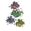
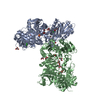
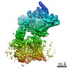
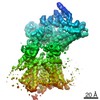
- PDB-1azs: COMPLEX OF GS-ALPHA WITH THE CATALYTIC DOMAINS OF MAMMALIAN ADENY... -
+ Open data

- Basic information
| Entry | Database: PDB / ID: 1azs | ||||||
|---|---|---|---|---|---|---|---|
| Title | COMPLEX OF GS-ALPHA WITH THE CATALYTIC DOMAINS OF MAMMALIAN ADENYLYL CYCLASE | ||||||
 Components Components |
| ||||||
 Keywords Keywords | COMPLEX (LYASE/HYDROLASE) / COMPLEX (LYASE-HYDROLASE) / HYDROLASE / SIGNAL TRANSDUCING PROTEIN / CYCLASE / EFFECTOR ENZYME / COMPLEX (LYASE-HYDROLASE) complex | ||||||
| Function / homology |  Function and homology information Function and homology informationAdenylate cyclase activating pathway / Hedgehog 'off' state / PKA activation / Adenylate cyclase inhibitory pathway / adenylate cyclase / sensory perception of chemical stimulus / regulation of insulin secretion involved in cellular response to glucose stimulus / mu-type opioid receptor binding / cAMP biosynthetic process / corticotropin-releasing hormone receptor 1 binding ...Adenylate cyclase activating pathway / Hedgehog 'off' state / PKA activation / Adenylate cyclase inhibitory pathway / adenylate cyclase / sensory perception of chemical stimulus / regulation of insulin secretion involved in cellular response to glucose stimulus / mu-type opioid receptor binding / cAMP biosynthetic process / corticotropin-releasing hormone receptor 1 binding / adenylate cyclase activity / G alpha (z) signalling events / beta-2 adrenergic receptor binding / adenylate cyclase binding / D1 dopamine receptor binding / adenylate cyclase-activating adrenergic receptor signaling pathway / insulin-like growth factor receptor binding / ionotropic glutamate receptor binding / cellular response to forskolin / adenylate cyclase activator activity / adenylate cyclase-modulating G protein-coupled receptor signaling pathway / G-protein beta/gamma-subunit complex binding / adenylate cyclase-activating G protein-coupled receptor signaling pathway / adenylate cyclase-activating dopamine receptor signaling pathway / manganese ion binding / heterotrimeric G-protein complex / positive regulation of cytosolic calcium ion concentration / Hydrolases; Acting on acid anhydrides; Acting on GTP to facilitate cellular and subcellular movement / intracellular signal transduction / cilium / membrane raft / GTPase activity / dendrite / GTP binding / magnesium ion binding / protein-containing complex / ATP binding / metal ion binding / membrane / plasma membrane / cytoplasm Similarity search - Function | ||||||
| Biological species |  | ||||||
| Method |  X-RAY DIFFRACTION / SYNCHROTRON / MOLECULAR REPLACEMENT / Resolution: 2.3 Å X-RAY DIFFRACTION / SYNCHROTRON / MOLECULAR REPLACEMENT / Resolution: 2.3 Å | ||||||
 Authors Authors | Tesmer, J.J.G. / Sprang, S.R. | ||||||
 Citation Citation | Journal: Science / Year: 1997 Title: Crystal structure of the catalytic domains of adenylyl cyclase in a complex with Gsalpha.GTPgammaS. Authors: Tesmer, J.J. / Sunahara, R.K. / Gilman, A.G. / Sprang, S.R. | ||||||
| History |
|
- Structure visualization
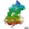
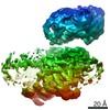
| Structure viewer | Molecule: MolmilJmol/JSmol |
|---|
- Downloads & links
-Download
| PDBx/mmCIF format | 1azs.cif.gz | 164.1 KB | Display | PDBx/mmCIF format |
|---|---|---|---|---|
| PDB format | pdb1azs.ent.gz | 124.9 KB | Display | PDB format |
| PDBx/mmJSON format | 1azs.json.gz | Tree view | PDBx/mmJSON format | |
| Others |  Other downloads Other downloads |
-Validation report
| Arichive directory | https://data.pdbj.org/pub/pdb/validation_reports/az/1azsftp://data.pdbj.org/pub/pdb/validation_reports/az/1azs | HTTPS FTP |
|---|
-Related structure data
| Related structure data |  1giaS S: Starting model for refinement |
|---|---|
| Similar structure data |


-Links
 PDBj
PDBj


- Assembly
| Deposited unit | 
| ||||||||
|---|---|---|---|---|---|---|---|---|---|
| 1 | 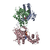
| ||||||||
| Unit cell |
|
-Components
-Protein , 3 types, 3 molecules ABC
| #1: Protein | Mass: 25010.822 Da / Num. of mol.: 1 / Fragment: C1A DOMAIN OF ADENYLYL CYCLASE / Mutation: V476M, N-TERMINAL HEXAHISTIDINE TAG Source method: isolated from a genetically manipulated source Source: (gene. exp.)  |
|---|---|
| #2: Protein | Mass: 23717.033 Da / Num. of mol.: 1 / Fragment: C2A DOMAIN OF ADENYLYL CYCLASE Source method: isolated from a genetically manipulated source Source: (gene. exp.) |
| #3: Protein | Mass: 46712.500 Da / Num. of mol.: 1 Mutation: C-TERMINAL HEXAHISTIDINE TAG, NOT PALMITOYLATED AT AMINO TERMINUS Source method: isolated from a genetically manipulated source Source: (gene. exp.) |
-Non-polymers , 4 types, 72 molecules 


| #4: Chemical | ChemComp-FKP /  Mass: 550.727 Da / Num. of mol.: 1 / Source method: obtained synthetically / Formula: C30H50N2O7 Mass: 550.727 Da / Num. of mol.: 1 / Source method: obtained synthetically / Formula: C30H50N2O7 |
|---|---|
| #5: Chemical | ChemComp-MG /  Mass: 24.305 Da / Num. of mol.: 1 / Source method: obtained synthetically / Formula: Mg Mass: 24.305 Da / Num. of mol.: 1 / Source method: obtained synthetically / Formula: Mg |
| #6: Chemical | ChemComp-GSP /  Mass: 539.246 Da / Num. of mol.: 1 / Source method: obtained synthetically / Formula: C10H16N5O13P3S Mass: 539.246 Da / Num. of mol.: 1 / Source method: obtained synthetically / Formula: C10H16N5O13P3S |
| #7: Water | ChemComp-HOH / Mass: 18.015 Da / Num. of mol.: 69 / Source method: isolated from a natural source / Formula: H2O |
-Experimental details
-Experiment
| Experiment | Method: X-RAY DIFFRACTION / Number of used crystals: 1 |
|---|
- Sample preparation
| Crystal | Density Matthews: 3 Å3/Da / Density % sol: 58 % Description: DUE TO THE ANISOTROPIC DIFFRACTION, DATA WITH |L| 18 WERE DISCARDED PRIOR TO SCALING. INCLUDING THIS DATA, COMPLETENESS IS 96.7%, REDUNDANCY IS 3.1, RSYM IS 11.6%, AND AVERAGE I/SIGMA(I) ...Description: DUE TO THE ANISOTROPIC DIFFRACTION, DATA WITH |L| 18 WERE DISCARDED PRIOR TO SCALING. INCLUDING THIS DATA, COMPLETENESS IS 96.7%, REDUNDANCY IS 3.1, RSYM IS 11.6%, AND AVERAGE I/SIGMA(I) IS 8.6. AVERAGE I/SIGMA (I) FOR THE OMITTED REFLECTIONS (AFTER SCALING) IS 1.7. | ||||||||||||||||||||||||||||||||||||||||||||||||||||||||
|---|---|---|---|---|---|---|---|---|---|---|---|---|---|---|---|---|---|---|---|---|---|---|---|---|---|---|---|---|---|---|---|---|---|---|---|---|---|---|---|---|---|---|---|---|---|---|---|---|---|---|---|---|---|---|---|---|---|
| Crystal grow | Method: vapor diffusion, hanging drop / pH: 5.6 Details: CRYSTALLIZED IN HANGING DROPS CONTAINING PROTEIN MIXED 1:1 WITH WELL SOLUTION OF 7.2-7.5% PEG 8000, 500MM NACL AND 100 MM (PH 5.4-5.6), vapor diffusion - hanging drop PH range: 5.4-5.6 | ||||||||||||||||||||||||||||||||||||||||||||||||||||||||
| Crystal | *PLUS | ||||||||||||||||||||||||||||||||||||||||||||||||||||||||
| Crystal grow | *PLUS Method: vapor diffusion / PH range low: 5.6 / PH range high: 5.4 | ||||||||||||||||||||||||||||||||||||||||||||||||||||||||
| Components of the solutions | *PLUS
|
-Data collection
| Diffraction | Mean temperature: 100 K |
|---|---|
| Diffraction source | Source: SYNCHROTRON / Site: CHESS  / Beamline: A1 / Wavelength: 0.908 / Beamline: A1 / Wavelength: 0.908 |
| Detector | Type: ADSC / Detector: CCD / Date: Jun 1, 1997 |
| Radiation | Monochromatic (M) / Laue (L): M / Scattering type: x-ray |
| Radiation wavelength | Wavelength: 0.908 Å / Relative weight: 1 |
| Reflection | Highest resolution: 2.3 Å / Num. obs: 37320 / % possible obs: 75.1 % / Observed criterion σ(I): -2 / Redundancy: 3.3 % / Biso Wilson estimate: 30.6 Å2 / Rsym value: 0.095 / Net I/σ(I): 11.5 |
| Reflection shell | Resolution: 2.3→2.34 Å / Redundancy: 2.8 % / Mean I/σ(I) obs: 2.5 / Rsym value: 0.333 / % possible all: 46.6 |
| Reflection | *PLUS Num. all: 48048 / Rmerge(I) obs: 0.095 |
- Processing
| Software |
| ||||||||||||||||||||||||||||||||||||||||||||||||||||||||||||||||||||||||||||||||
|---|---|---|---|---|---|---|---|---|---|---|---|---|---|---|---|---|---|---|---|---|---|---|---|---|---|---|---|---|---|---|---|---|---|---|---|---|---|---|---|---|---|---|---|---|---|---|---|---|---|---|---|---|---|---|---|---|---|---|---|---|---|---|---|---|---|---|---|---|---|---|---|---|---|---|---|---|---|---|---|---|---|
| Refinement | Method to determine structure: MOLECULAR REPLACEMENT Starting model: PDB ENTRY 1GIA Resolution: 2.3→15 Å / Rfactor Rfree error: 0.005 / Isotropic thermal model: RESTRAINED / Cross valid method: THROUGHOUT / Details: A BULK SOLVENT CORRECTION WAS USED.
| ||||||||||||||||||||||||||||||||||||||||||||||||||||||||||||||||||||||||||||||||
| Displacement parameters | Biso mean: 52.2 Å2
| ||||||||||||||||||||||||||||||||||||||||||||||||||||||||||||||||||||||||||||||||
| Refine analyze | Luzzati d res low obs: 15 Å / Luzzati sigma a obs: 0.31 Å | ||||||||||||||||||||||||||||||||||||||||||||||||||||||||||||||||||||||||||||||||
| Refinement step | Cycle: LAST / Resolution: 2.3→15 Å
| ||||||||||||||||||||||||||||||||||||||||||||||||||||||||||||||||||||||||||||||||
| Refine LS restraints |
| ||||||||||||||||||||||||||||||||||||||||||||||||||||||||||||||||||||||||||||||||
| LS refinement shell | Resolution: 2.3→2.4 Å / Rfactor Rfree error: 0.022 / Total num. of bins used: 8
| ||||||||||||||||||||||||||||||||||||||||||||||||||||||||||||||||||||||||||||||||
| Xplor file |
|
