 Movie
Movie Controller

[English] 日本語
 Yorodumi
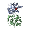
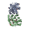
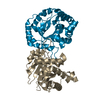
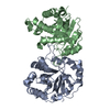
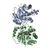
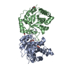
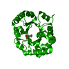
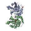
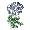
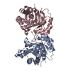
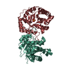
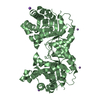
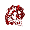
- PDB-1ag1: MONOHYDROGEN PHOSPHATE BINDING TO TRYPANOSOMAL TRIOSEPHOSPHATE IS... -
+ Open data

- Basic information
| Entry | Database: PDB / ID: 1ag1 | ||||||
|---|---|---|---|---|---|---|---|
| Title | MONOHYDROGEN PHOSPHATE BINDING TO TRYPANOSOMAL TRIOSEPHOSPHATE ISOMERASE | ||||||
 Components Components | TRIOSEPHOSPHATE ISOMERASE | ||||||
 Keywords Keywords | ISOMERASE(INTRAMOLECULAR OXIDOREDUCTASE) / ISOMERASE (INTRAMOLECULAR OXIDOREDUCTASE) | ||||||
| Function / homology |  Function and homology information Function and homology informationglycosome / triose-phosphate isomerase / triose-phosphate isomerase activity / glyceraldehyde-3-phosphate biosynthetic process / glycerol catabolic process / gluconeogenesis / glycolytic process / cytoplasm / cytosol Similarity search - Function | ||||||
| Biological species |  | ||||||
| Method |  X-RAY DIFFRACTION / MOLECULAR REPLACEMENT / Resolution: 2.36 Å X-RAY DIFFRACTION / MOLECULAR REPLACEMENT / Resolution: 2.36 Å | ||||||
 Authors Authors | Verlinde, C.L.M.J. / Hol, W.G.J. | ||||||
 Citation Citation | Journal: Eur.J.Biochem. / Year: 1991 Title: Anion binding at the active site of trypanosomal triosephosphate isomerase. Monohydrogen phosphate does not mimic sulphate. Authors: Verlinde, C.L. / Noble, M.E. / Kalk, K.H. / Groendijk, H. / Wierenga, R.K. / Hol, W.G. | ||||||
| History |
|
- Structure visualization
| Structure viewer | Molecule: MolmilJmol/JSmol |
|---|
- Downloads & links
-Download
| PDBx/mmCIF format | 1ag1.cif.gz | 106.2 KB | Display | PDBx/mmCIF format |
|---|---|---|---|---|
| PDB format | pdb1ag1.ent.gz | 82.4 KB | Display | PDB format |
| PDBx/mmJSON format | 1ag1.json.gz | Tree view | PDBx/mmJSON format | |
| Others |  Other downloads Other downloads |
-Validation report
| Summary document | 1ag1_validation.pdf.gz | 392.4 KB | Display | wwPDB validaton report |
|---|---|---|---|---|
| Full document | 1ag1_full_validation.pdf.gz | 412.1 KB | Display | |
| Data in XML | 1ag1_validation.xml.gz | 13.4 KB | Display | |
| Data in CIF | 1ag1_validation.cif.gz | 20.6 KB | Display | |
| Arichive directory | https://data.pdbj.org/pub/pdb/validation_reports/ag/1ag1ftp://data.pdbj.org/pub/pdb/validation_reports/ag/1ag1 | HTTPS FTP |
-Related structure data
| Related structure data | 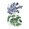 6timS S: Starting model for refinement |
|---|---|
| Similar structure data |


-Links
 PDBj
PDBj
- Assembly
| Deposited unit | 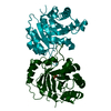
| ||||||||
|---|---|---|---|---|---|---|---|---|---|
| 1 |
| ||||||||
| Unit cell |
|
-Components
| #1: Protein | Mass: 26865.832 Da / Num. of mol.: 2 / Source method: isolated from a natural source / Source: (natural) #2: Chemical | ChemComp-PO4 / |   Mass: 94.971 Da / Num. of mol.: 1 / Source method: obtained synthetically / Formula: PO4 Mass: 94.971 Da / Num. of mol.: 1 / Source method: obtained synthetically / Formula: PO4#3: Water | ChemComp-HOH / |  Mass: 18.015 Da / Num. of mol.: 156 / Source method: isolated from a natural source / Formula: H2O Mass: 18.015 Da / Num. of mol.: 156 / Source method: isolated from a natural source / Formula: H2O |
|---|
-Experimental details
-Experiment
| Experiment | Method: X-RAY DIFFRACTION / Number of used crystals: 1 |
|---|
- Sample preparation
| Crystal | Density Matthews: 2.39 Å3/Da / Density % sol: 48.59 % | |||||||||||||||||||||||||||||||||||||||||||||
|---|---|---|---|---|---|---|---|---|---|---|---|---|---|---|---|---|---|---|---|---|---|---|---|---|---|---|---|---|---|---|---|---|---|---|---|---|---|---|---|---|---|---|---|---|---|---|
| Crystal grow | pH: 7 Details: 2.4 M AMMONIUM SULFATE IN 0.2 MOPS BUFFER, PH 7.0 FOLLOWED BY TRANSFER TO 44% PEG-6000 CONTAINING 15 MM PHOSPHATE | |||||||||||||||||||||||||||||||||||||||||||||
| Crystal grow | *PLUS pH: 7 / Method: vapor diffusion, hanging drop / Details: Wierenga, R.K., (1984) J. Mol. Biol., 178, 487. | |||||||||||||||||||||||||||||||||||||||||||||
| Components of the solutions | *PLUS
|
-Data collection
| Diffraction | Mean temperature: 298 K |
|---|---|
| Diffraction source | Source: ROTATING ANODE / Type: ELLIOTT GX-21 / Wavelength: 1.5418 |
| Detector | Type: ENRAF-NONIUS FAST / Detector: DIFFRACTOMETER / Date: Mar 1, 1990 / Details: STANDARD |
| Radiation | Monochromator: GRAPHITE(002) / Monochromatic (M) / Laue (L): M / Scattering type: x-ray |
| Radiation wavelength | Wavelength: 1.5418 Å / Relative weight: 1 |
| Reflection | Highest resolution: 2.36 Å / Num. obs: 17592 / % possible obs: 87.5 % / Observed criterion σ(I): 2 / Redundancy: 1.7 % / Rmerge(I) obs: 0.063 |
- Processing
| Software |
| ||||||||||||||||||||||||||||||
|---|---|---|---|---|---|---|---|---|---|---|---|---|---|---|---|---|---|---|---|---|---|---|---|---|---|---|---|---|---|---|---|
| Refinement | Method to determine structure: MOLECULAR REPLACEMENT Starting model: PDB ENTRY 6TIM Resolution: 2.36→6 Å / Isotropic thermal model: TNT BCORREL V1.0 / Cross valid method: A POSTERIORI / σ(F): 3 / Stereochemistry target values: TNT PROTGEO /
| ||||||||||||||||||||||||||||||
| Refinement step | Cycle: LAST / Resolution: 2.36→6 Å
| ||||||||||||||||||||||||||||||
| Refine LS restraints |
| ||||||||||||||||||||||||||||||
| Software | *PLUS Name: TNT / Version: 5C / Classification: refinement | ||||||||||||||||||||||||||||||
| Refinement | *PLUS Rfactor obs: 0.15 / Rfactor Rwork: 0.15 | ||||||||||||||||||||||||||||||
| Solvent computation | *PLUS | ||||||||||||||||||||||||||||||
| Displacement parameters | *PLUS |
