 ムービー
ムービー コントローラー


+ データを開く

- 基本情報
基本情報
| 登録情報 | データベース: PDB / ID: 7rxd | ||||||
|---|---|---|---|---|---|---|---|
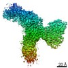
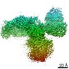
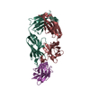
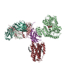
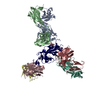
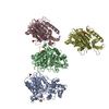
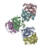
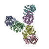
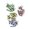
| タイトル | CryoEM structure of RBD domain of COVID-19 in complex with Legobody | ||||||
 要素 要素 |
| ||||||
 キーワード キーワード |  STRUCTURAL PROTEIN (タンパク質) / RBD / maltose-binding protein / Fab / nanobody (ナノボディ) STRUCTURAL PROTEIN (タンパク質) / RBD / maltose-binding protein / Fab / nanobody (ナノボディ) | ||||||
| 機能・相同性 |  機能・相同性情報 機能・相同性情報IgG binding / carbohydrate transmembrane transporter activity / Maturation of spike protein / viral translation / Translation of Structural Proteins / Virion Assembly and Release / host cell surface / host extracellular space / suppression by virus of host tetherin activity / Induction of Cell-Cell Fusion ...IgG binding / carbohydrate transmembrane transporter activity / Maturation of spike protein / viral translation / Translation of Structural Proteins / Virion Assembly and Release / host cell surface / host extracellular space / suppression by virus of host tetherin activity / Induction of Cell-Cell Fusion / structural constituent of virion / host cell endoplasmic reticulum-Golgi intermediate compartment membrane / entry receptor-mediated virion attachment to host cell / receptor-mediated endocytosis of virus by host cell / Attachment and Entry / membrane fusion / positive regulation of viral entry into host cell / receptor-mediated virion attachment to host cell / receptor ligand activity / ペリプラズム / host cell surface receptor binding / fusion of virus membrane with host plasma membrane / fusion of virus membrane with host endosome membrane / エンベロープ (ウイルス) / symbiont-mediated suppression of host type I interferon-mediated signaling pathway / virion attachment to host cell / SARS-CoV-2 activates/modulates innate and adaptive immune responses / host cell plasma membrane / virion membrane / extracellular region / 生体膜 / identical protein binding / 細胞膜類似検索 - 分子機能 | ||||||
| 生物種 |  Escherichia coli (大腸菌) Escherichia coli (大腸菌) Staphylococcus aureus (黄色ブドウ球菌)Streptococcus sp. (バクテリア) Staphylococcus aureus (黄色ブドウ球菌)Streptococcus sp. (バクテリア) Mus musculus (ハツカネズミ)Vicugna pacos (アルパカ) Mus musculus (ハツカネズミ)Vicugna pacos (アルパカ)  Severe acute respiratory syndrome coronavirus 2 (SARSコロナウイルス2) Severe acute respiratory syndrome coronavirus 2 (SARSコロナウイルス2) | ||||||
| 手法 | 電子顕微鏡法 / 単粒子再構成法 / クライオ電子顕微鏡法 / 解像度: 3.6 Å | ||||||
 データ登録者 データ登録者 | Wu, X.D. / Rapoport, T.A. | ||||||
| 資金援助 |  米国, 1件 米国, 1件
| ||||||
 引用 引用 | ジャーナル: Proc Natl Acad Sci U S A / 年: 2021 タイトル: Cryo-EM structure determination of small proteins by nanobody-binding scaffolds (Legobodies). 著者: Xudong Wu / Tom A Rapoport / 要旨: We describe a general method that allows structure determination of small proteins by single-particle cryo-electron microscopy (cryo-EM). The method is based on the availability of a target-binding ...We describe a general method that allows structure determination of small proteins by single-particle cryo-electron microscopy (cryo-EM). The method is based on the availability of a target-binding nanobody, which is then rigidly attached to two scaffolds: 1) a Fab fragment of an antibody directed against the nanobody and 2) a nanobody-binding protein A fragment fused to maltose binding protein and Fab-binding domains. The overall ensemble of ∼120 kDa, called Legobody, does not perturb the nanobody-target interaction, is easily recognizable in EM images due to its unique shape, and facilitates particle alignment in cryo-EM image processing. The utility of the method is demonstrated for the KDEL receptor, a 23-kDa membrane protein, resulting in a map at 3.2-Å overall resolution with density sufficient for de novo model building, and for the 22-kDa receptor-binding domain (RBD) of SARS-CoV-2 spike protein, resulting in a map at 3.6-Å resolution that allows analysis of the binding interface to the nanobody. The Legobody approach thus overcomes the current size limitations of cryo-EM analysis. | ||||||
| 履歴 |
|
- 構造の表示
構造の表示
| ムービー |
ムービービューア |
|---|---|
| 構造ビューア | 分子: MolmilJmol/JSmol |
- ダウンロードとリンク
-ダウンロード
| PDBx/mmCIF形式 | 7rxd.cif.gz | 211.3 KB | 表示 | PDBx/mmCIF形式 |
|---|---|---|---|---|
| PDB形式 | pdb7rxd.ent.gz | 170.5 KB | 表示 | PDB形式 |
| PDBx/mmJSON形式 | 7rxd.json.gz | ツリー表示 | PDBx/mmJSON形式 | |
| その他 |  その他のダウンロード その他のダウンロード |
-検証レポート
| アーカイブディレクトリ | https://data.pdbj.org/pub/pdb/validation_reports/rx/7rxdftp://data.pdbj.org/pub/pdb/validation_reports/rx/7rxd | HTTPS FTP |
|---|
-関連構造データ


-リンク
 PDBj
PDBj


- 集合体
集合体
| 登録構造単位 | 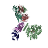
|
|---|---|
| 1 |
|
-要素
-抗体 , 4種, 4分子 BHLN
| #1: 抗体 | 分子量: 59233.246 Da / 分子数: 1 / 由来タイプ: 組換発現 由来: (組換発現) Escherichia coli (大腸菌), (組換発現) Staphylococcus aureus (黄色ブドウ球菌), (組換発現) Streptococcus sp. (バクテリア)遺伝子: DAH37_23060, spa, spg / 発現宿主: Escherichia coli (大腸菌)参照: UniProt: A0A4Z0THX4, UniProt: P99134, UniProt: P06654 |
|---|---|
| #2: 抗体 | 分子量: 25252.217 Da / 分子数: 1 / 由来タイプ: 組換発現 / 由来: (組換発現) Mus musculus (ハツカネズミ) / 発現宿主:  Homo sapiens (ヒト) Homo sapiens (ヒト) |
| #3: 抗体 | 分子量: 24095.852 Da / 分子数: 1 / 由来タイプ: 組換発現 / 由来: (組換発現) Mus musculus (ハツカネズミ) / 発現宿主: Homo sapiens (ヒト) |
| #4: 抗体 | 分子量: 14661.201 Da / 分子数: 1 / 由来タイプ: 組換発現 / 由来: (組換発現) Vicugna pacos (アルパカ) / 発現宿主: Escherichia coli (大腸菌) |
-タンパク質 / 糖 , 2種, 2分子 R
| #5: タンパク質 | 分子量: 25995.555 Da / 分子数: 1 / Fragment: Receptor Binding Domain (RBD) / 由来タイプ: 組換発現 由来: (組換発現) Severe acute respiratory syndrome coronavirus 2 (SARSコロナウイルス2)遺伝子: S, 2 / 発現宿主: Homo sapiens (ヒト) / 参照: UniProt: P0DTC2 |
|---|---|
| #6: 多糖 | alpha-D-glucopyranose-(1-4)-alpha-D-glucopyranose  オリゴ糖, Oligosaccharideオリゴ糖 オリゴ糖, Oligosaccharideオリゴ糖クラス: 栄養素 栄養素 / 分子量: 342.297 Da / 分子数: 1 / 由来タイプ: 組換発現 / 詳細: oligosaccharide / 参照: alpha-maltose |
-詳細
| 研究の焦点であるリガンドがあるか | N |
|---|
-実験情報
-実験
| 実験 | 手法: 電子顕微鏡法 |
|---|---|
| EM実験 | 試料の集合状態: PARTICLE / 3次元再構成法: 単粒子再構成法 |
- 試料調製
試料調製
| 構成要素 | 名称: The complex of RBD domain of COVID-19 with Legobody / タイプ: COMPLEX / Entity ID: #1-#5 / 由来: MULTIPLE SOURCES |
|---|---|
| 分子量 | 値: 0.15 MDa / 実験値: NO |
| 緩衝液 | pH: 7.4 |
| 試料 | 包埋: NO / シャドウイング: NO / 染色: NO / 凍結: YES |
| 急速凍結 | 凍結剤: ETHANE |
- 電子顕微鏡撮影
| 実験機器 |  モデル: Titan Krios / 画像提供: FEI Company |
|---|---|
| 顕微鏡 | モデル: FEI TITAN KRIOS |
| 電子銃 | 電子線源: FIELD EMISSION GUN / 加速電圧: 300 kV / 照射モード: FLOOD BEAM |
| 電子レンズ | モード: BRIGHT FIELDBright-field microscopy |
| 撮影 | 電子線照射量: 50.42 e/Å2 / フィルム・検出器のモデル: GATAN K3 (6k x 4k) |
- 解析
解析
| ソフトウェア | 名称: PHENIX / バージョン: 1.19.1_4122: / 分類: 精密化 | ||||||||||||||||||||||||
|---|---|---|---|---|---|---|---|---|---|---|---|---|---|---|---|---|---|---|---|---|---|---|---|---|---|
| CTF補正 | タイプ: PHASE FLIPPING AND AMPLITUDE CORRECTION | ||||||||||||||||||||||||
| 3次元再構成 | 解像度: 3.6 Å / 解像度の算出法: FSC 0.143 CUT-OFF / 粒子像の数: 282995 / 対称性のタイプ: POINT | ||||||||||||||||||||||||
| 拘束条件 |
|
