 Movie
Movie Controller

[English] 日本語
 Yorodumi
- PDB-7r6x: SARS-CoV-2 spike receptor-binding domain (RBD) in complex with S2... -
+ Open data

- Basic information
| Entry | Database: PDB / ID: 7r6x | ||||||
|---|---|---|---|---|---|---|---|
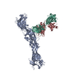
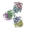
| Title | SARS-CoV-2 spike receptor-binding domain (RBD) in complex with S2E12 Fab, S309 Fab, and S304 Fab | ||||||
 Components Components |
| ||||||
 Keywords Keywords | VIRAL PROTEIN/IMMUNE SYSTEM / COVID-19 / SARS-CoV-2 / neutralizing monoclonal antibody / VIRAL PROTEIN-IMMUNE SYSTEM complex | ||||||
| Function / homology |  Function and homology information Function and homology informationsymbiont-mediated disruption of host tissue / Maturation of spike protein / Translation of Structural Proteins / Virion Assembly and Release / host cell surface / host extracellular region / symbiont-mediated-mediated suppression of host tetherin activity / Induction of Cell-Cell Fusion / structural constituent of virion / positive regulation of viral entry into host cell ...symbiont-mediated disruption of host tissue / Maturation of spike protein / Translation of Structural Proteins / Virion Assembly and Release / host cell surface / host extracellular region / symbiont-mediated-mediated suppression of host tetherin activity / Induction of Cell-Cell Fusion / structural constituent of virion / positive regulation of viral entry into host cell / membrane fusion / host cell endoplasmic reticulum-Golgi intermediate compartment membrane / Attachment and Entry / entry receptor-mediated virion attachment to host cell / receptor-mediated virion attachment to host cell / host cell surface receptor binding / symbiont-mediated suppression of host innate immune response / endocytosis involved in viral entry into host cell / receptor ligand activity / fusion of virus membrane with host plasma membrane / fusion of virus membrane with host endosome membrane / viral envelope / symbiont entry into host cell / virion attachment to host cell / host cell plasma membrane / SARS-CoV-2 activates/modulates innate and adaptive immune responses / virion membrane / membrane / identical protein binding / plasma membrane Similarity search - Function | ||||||
| Biological species |  Homo sapiens (human) Homo sapiens (human)  Severe acute respiratory syndrome coronavirus 2 Severe acute respiratory syndrome coronavirus 2 | ||||||
| Method |  X-RAY DIFFRACTION / SYNCHROTRON / MOLECULAR REPLACEMENT / Resolution: 2.95 Å X-RAY DIFFRACTION / SYNCHROTRON / MOLECULAR REPLACEMENT / Resolution: 2.95 Å | ||||||
 Authors Authors | Snell, G. / Czudnochowski, N. / Croll, T.I. / Nix, J.C. / Corti, D. / Cameroni, E. / Pinto, D. / Beltramello, M. | ||||||
 Citation Citation | Journal: Nature / Year: 2021 Title: SARS-CoV-2 RBD antibodies that maximize breadth and resistance to escape. Authors: Tyler N Starr / Nadine Czudnochowski / Zhuoming Liu / Fabrizia Zatta / Young-Jun Park / Amin Addetia / Dora Pinto / Martina Beltramello / Patrick Hernandez / Allison J Greaney / Roberta ...Authors: Tyler N Starr / Nadine Czudnochowski / Zhuoming Liu / Fabrizia Zatta / Young-Jun Park / Amin Addetia / Dora Pinto / Martina Beltramello / Patrick Hernandez / Allison J Greaney / Roberta Marzi / William G Glass / Ivy Zhang / Adam S Dingens / John E Bowen / M Alejandra Tortorici / Alexandra C Walls / Jason A Wojcechowskyj / Anna De Marco / Laura E Rosen / Jiayi Zhou / Martin Montiel-Ruiz / Hannah Kaiser / Josh R Dillen / Heather Tucker / Jessica Bassi / Chiara Silacci-Fregni / Michael P Housley / Julia di Iulio / Gloria Lombardo / Maria Agostini / Nicole Sprugasci / Katja Culap / Stefano Jaconi / Marcel Meury / Exequiel Dellota / Rana Abdelnabi / Shi-Yan Caroline Foo / Elisabetta Cameroni / Spencer Stumpf / Tristan I Croll / Jay C Nix / Colin Havenar-Daughton / Luca Piccoli / Fabio Benigni / Johan Neyts / Amalio Telenti / Florian A Lempp / Matteo S Pizzuto / John D Chodera / Christy M Hebner / Herbert W Virgin / Sean P J Whelan / David Veesler / Davide Corti / Jesse D Bloom / Gyorgy Snell /     Abstract: An ideal therapeutic anti-SARS-CoV-2 antibody would resist viral escape, have activity against diverse sarbecoviruses, and be highly protective through viral neutralization and effector functions. ...An ideal therapeutic anti-SARS-CoV-2 antibody would resist viral escape, have activity against diverse sarbecoviruses, and be highly protective through viral neutralization and effector functions. Understanding how these properties relate to each other and vary across epitopes would aid the development of therapeutic antibodies and guide vaccine design. Here we comprehensively characterize escape, breadth and potency across a panel of SARS-CoV-2 antibodies targeting the receptor-binding domain (RBD). Despite a trade-off between in vitro neutralization potency and breadth of sarbecovirus binding, we identify neutralizing antibodies with exceptional sarbecovirus breadth and a corresponding resistance to SARS-CoV-2 escape. One of these antibodies, S2H97, binds with high affinity across all sarbecovirus clades to a cryptic epitope and prophylactically protects hamsters from viral challenge. Antibodies that target the angiotensin-converting enzyme 2 (ACE2) receptor-binding motif (RBM) typically have poor breadth and are readily escaped by mutations despite high neutralization potency. Nevertheless, we also characterize a potent RBM antibody (S2E12) with breadth across sarbecoviruses related to SARS-CoV-2 and a high barrier to viral escape. These data highlight principles underlying variation in escape, breadth and potency among antibodies that target the RBD, and identify epitopes and features to prioritize for therapeutic development against the current and potential future pandemics. #1: Journal: To Be PublishedTitle: SARS-CoV-2 RBD antibodies that maximize breadth and resistance to escape Authors: Starr, T.N. / Czudnochowski, N. / Liu, Z. / Zatta, F. / Park, Y.J. / Pinto, D. / Beltramello, M. / Hernandez, P. / Cameroni, E. / Croll, T.I. / Nix, J.C. / Chodera, J.D. / Whelan, S.P.J. / ...Authors: Starr, T.N. / Czudnochowski, N. / Liu, Z. / Zatta, F. / Park, Y.J. / Pinto, D. / Beltramello, M. / Hernandez, P. / Cameroni, E. / Croll, T.I. / Nix, J.C. / Chodera, J.D. / Whelan, S.P.J. / Virgin, H.W. / Corti, D. / Veesler, D. / Bloom, J.D. / Snell, G. | ||||||
| History |
|
- Structure visualization
| Structure viewer | Molecule: MolmilJmol/JSmol |
|---|
- Downloads & links
-Download
| PDBx/mmCIF format | 7r6x.cif.gz | 483.3 KB | Display | PDBx/mmCIF format |
|---|---|---|---|---|
| PDB format | pdb7r6x.ent.gz | 394.7 KB | Display | PDB format |
| PDBx/mmJSON format | 7r6x.json.gz | Tree view | PDBx/mmJSON format | |
| Others |  Other downloads Other downloads |
-Validation report
| Arichive directory | https://data.pdbj.org/pub/pdb/validation_reports/r6/7r6xftp://data.pdbj.org/pub/pdb/validation_reports/r6/7r6x | HTTPS FTP |
|---|
-Related structure data
| Related structure data |  7m7wC  7r6wC  7r7nC  7jx3S  7k3qS C: citing same article ( S: Starting model for refinement |
|---|---|
| Similar structure data |


-Links
 PDBj
PDBj


- Assembly
| Deposited unit | 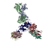
| ||||||||
|---|---|---|---|---|---|---|---|---|---|
| 1 |
| ||||||||
| Unit cell |
|
-Components
-Antibody , 6 types, 6 molecules LHBADC
| #1: Antibody | Mass: 23369.947 Da / Num. of mol.: 1 Source method: isolated from a genetically manipulated source Source: (gene. exp.) Homo sapiens (human) / Cell line (production host): HEK293.sus / Production host: Homo sapiens (human) |
|---|---|
| #2: Antibody | Mass: 23729.389 Da / Num. of mol.: 1 Source method: isolated from a genetically manipulated source Source: (gene. exp.) Homo sapiens (human) / Cell line (production host): HEK293.sus / Production host: Homo sapiens (human) |
| #4: Antibody | Mass: 23204.697 Da / Num. of mol.: 1 Source method: isolated from a genetically manipulated source Source: (gene. exp.) Homo sapiens (human) / Cell line (production host): HEK293.sus / Production host: Homo sapiens (human) |
| #5: Antibody | Mass: 24573.471 Da / Num. of mol.: 1 Source method: isolated from a genetically manipulated source Source: (gene. exp.) Homo sapiens (human) / Cell line (production host): HEK293.sus / Production host: Homo sapiens (human) |
| #6: Antibody | Mass: 23546.127 Da / Num. of mol.: 1 Source method: isolated from a genetically manipulated source Source: (gene. exp.) Homo sapiens (human) / Production host:   Cricetulus griseus (Chinese hamster) Cricetulus griseus (Chinese hamster) |
| #7: Antibody | Mass: 24016.900 Da / Num. of mol.: 1 Source method: isolated from a genetically manipulated source Source: (gene. exp.) Homo sapiens (human) / Production host: Cricetulus griseus (Chinese hamster) |
-Protein / Sugars , 2 types, 2 molecules R
| #3: Protein | Mass: 24442.332 Da / Num. of mol.: 1 / Fragment: receptor-binding domain (UNP residues 328-531) Source method: isolated from a genetically manipulated source Source: (gene. exp.) Severe acute respiratory syndrome coronavirus 2Gene: S, 2 / Production host: Homo sapiens (human) / References: UniProt: P0DTC2 |
|---|---|
| #8: Sugar | ChemComp-NAG /  Type: D-saccharide, beta linking / Mass: 221.208 Da / Num. of mol.: 1 / Source method: obtained synthetically / Formula: C8H15NO6 Type: D-saccharide, beta linking / Mass: 221.208 Da / Num. of mol.: 1 / Source method: obtained synthetically / Formula: C8H15NO6 |
-Non-polymers , 2 types, 5 molecules 

| #9: Chemical |  Mass: 35.453 Da / Num. of mol.: 2 / Source method: obtained synthetically / Formula: Cl Mass: 35.453 Da / Num. of mol.: 2 / Source method: obtained synthetically / Formula: Cl#10: Water | ChemComp-HOH / | Mass: 18.015 Da / Num. of mol.: 3 / Source method: isolated from a natural source / Formula: H2O |
|---|
-Details
| Has ligand of interest | N |
|---|---|
| Has protein modification | Y |
-Experimental details
-Experiment
| Experiment | Method: X-RAY DIFFRACTION / Number of used crystals: 1 |
|---|
- Sample preparation
| Crystal | Density Matthews: 5.37 Å3/Da / Density % sol: 77.11 % |
|---|---|
| Crystal grow | Temperature: 293.15 K / Method: vapor diffusion, sitting drop Details: Crystal 1: 0.09 M phosphate/citrate, pH 5.5, 27% v/v PEG Smear Low, 4% v/v polypropylene glycol 400, 0.02 M imidazole, pH 7, Crystal 2: 0.09 M phosphate/citrate, pH 5.5, 27% v/v PEG Smear ...Details: Crystal 1: 0.09 M phosphate/citrate, pH 5.5, 27% v/v PEG Smear Low, 4% v/v polypropylene glycol 400, 0.02 M imidazole, pH 7, Crystal 2: 0.09 M phosphate/citrate, pH 5.5, 27% v/v PEG Smear Low, 0.01 M potassium/sodium phosphate, pH 7, 1% v/v PPGBA 230, 1.5% v/v PPGBA 400 |
-Data collection
| Diffraction | Mean temperature: 100 K / Serial crystal experiment: N | ||||||||||||||||||||||||||||||
|---|---|---|---|---|---|---|---|---|---|---|---|---|---|---|---|---|---|---|---|---|---|---|---|---|---|---|---|---|---|---|---|
| Diffraction source | Source: SYNCHROTRON / Site: ALS / Beamline: 4.2.2 / Wavelength: 1.00003 Å | ||||||||||||||||||||||||||||||
| Detector | Type: RDI CMOS_8M / Detector: CMOS / Date: Oct 4, 2020 | ||||||||||||||||||||||||||||||
| Radiation | Protocol: SINGLE WAVELENGTH / Monochromatic (M) / Laue (L): M / Scattering type: x-ray | ||||||||||||||||||||||||||||||
| Radiation wavelength | Wavelength: 1.00003 Å / Relative weight: 1 | ||||||||||||||||||||||||||||||
| Reflection | Resolution: 2.93→49 Å / Num. obs: 77621 / % possible obs: 100 % / Redundancy: 28.9 % / CC1/2: 0.998 / Rmerge(I) obs: 0.295 / Rpim(I) all: 0.056 / Rrim(I) all: 0.3 / Net I/σ(I): 13.3 / Num. measured all: 2242103 | ||||||||||||||||||||||||||||||
| Reflection shell | Diffraction-ID: 1
|
- Processing
| Software |
| ||||||||||||||||||||||||||||||||||||||||||||||||||||||||||||||||||||||||||||||||||||||||||||||||||||||||||||||||||||||||||||||||||||||||||||||||||||||||||||||||||||||||||||||||||||||||||||||||||||||||
|---|---|---|---|---|---|---|---|---|---|---|---|---|---|---|---|---|---|---|---|---|---|---|---|---|---|---|---|---|---|---|---|---|---|---|---|---|---|---|---|---|---|---|---|---|---|---|---|---|---|---|---|---|---|---|---|---|---|---|---|---|---|---|---|---|---|---|---|---|---|---|---|---|---|---|---|---|---|---|---|---|---|---|---|---|---|---|---|---|---|---|---|---|---|---|---|---|---|---|---|---|---|---|---|---|---|---|---|---|---|---|---|---|---|---|---|---|---|---|---|---|---|---|---|---|---|---|---|---|---|---|---|---|---|---|---|---|---|---|---|---|---|---|---|---|---|---|---|---|---|---|---|---|---|---|---|---|---|---|---|---|---|---|---|---|---|---|---|---|---|---|---|---|---|---|---|---|---|---|---|---|---|---|---|---|---|---|---|---|---|---|---|---|---|---|---|---|---|---|---|---|---|
| Refinement | Method to determine structure: MOLECULAR REPLACEMENT Starting model: PDB entries 7JX3 and 7K3Q Resolution: 2.95→40 Å / Cor.coef. Fo:Fc: 0.922 / Cor.coef. Fo:Fc free: 0.906 / SU B: 33.532 / SU ML: 0.26 / Cross valid method: THROUGHOUT / σ(F): 0 / ESU R: 0.357 / ESU R Free: 0.282 / Stereochemistry target values: MAXIMUM LIKELIHOOD / Details: U VALUES : WITH TLS ADDED
| ||||||||||||||||||||||||||||||||||||||||||||||||||||||||||||||||||||||||||||||||||||||||||||||||||||||||||||||||||||||||||||||||||||||||||||||||||||||||||||||||||||||||||||||||||||||||||||||||||||||||
| Solvent computation | Ion probe radii: 0.8 Å / Shrinkage radii: 0.8 Å / VDW probe radii: 1.2 Å / Solvent model: MASK | ||||||||||||||||||||||||||||||||||||||||||||||||||||||||||||||||||||||||||||||||||||||||||||||||||||||||||||||||||||||||||||||||||||||||||||||||||||||||||||||||||||||||||||||||||||||||||||||||||||||||
| Displacement parameters | Biso max: 240.68 Å2 / Biso mean: 116.456 Å2 / Biso min: 58.09 Å2
| ||||||||||||||||||||||||||||||||||||||||||||||||||||||||||||||||||||||||||||||||||||||||||||||||||||||||||||||||||||||||||||||||||||||||||||||||||||||||||||||||||||||||||||||||||||||||||||||||||||||||
| Refinement step | Cycle: final / Resolution: 2.95→40 Å
| ||||||||||||||||||||||||||||||||||||||||||||||||||||||||||||||||||||||||||||||||||||||||||||||||||||||||||||||||||||||||||||||||||||||||||||||||||||||||||||||||||||||||||||||||||||||||||||||||||||||||
| Refine LS restraints |
| ||||||||||||||||||||||||||||||||||||||||||||||||||||||||||||||||||||||||||||||||||||||||||||||||||||||||||||||||||||||||||||||||||||||||||||||||||||||||||||||||||||||||||||||||||||||||||||||||||||||||
| LS refinement shell | Resolution: 2.95→3.026 Å / Rfactor Rfree error: 0 / Total num. of bins used: 20
| ||||||||||||||||||||||||||||||||||||||||||||||||||||||||||||||||||||||||||||||||||||||||||||||||||||||||||||||||||||||||||||||||||||||||||||||||||||||||||||||||||||||||||||||||||||||||||||||||||||||||
| Refinement TLS params. | Method: refined / Refine-ID: X-RAY DIFFRACTION
| ||||||||||||||||||||||||||||||||||||||||||||||||||||||||||||||||||||||||||||||||||||||||||||||||||||||||||||||||||||||||||||||||||||||||||||||||||||||||||||||||||||||||||||||||||||||||||||||||||||||||
| Refinement TLS group |
|
