 Movie
Movie Controller


+ Open data

- Basic information
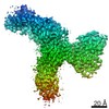
| Entry | Database: EMDB / ID: EMD-24728 | |||||||||
|---|---|---|---|---|---|---|---|---|---|---|
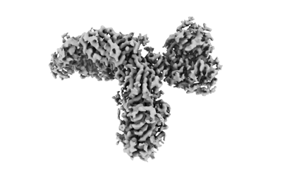
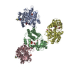
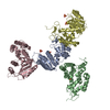
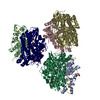
| Title | CryoEM structure of KDELR with Legobody | |||||||||
 Map data Map data | sharpened map for the complex | |||||||||
 Sample Sample |
| |||||||||
 Keywords Keywords | protein A / maltose-binding protein / Fab / nanobody / STRUCTURAL PROTEIN | |||||||||
| Function / homology |  Function and homology information Function and homology informationKDEL sequence binding / COPI-coated vesicle membrane / ER lumen protein retrieval receptor activity / COPI-dependent Golgi-to-ER retrograde traffic / COPI-mediated anterograde transport / protein retention in ER lumen / maintenance of protein localization in endoplasmic reticulum / cis-Golgi network / retrograde vesicle-mediated transport, Golgi to endoplasmic reticulum / IgG binding ...KDEL sequence binding / COPI-coated vesicle membrane / ER lumen protein retrieval receptor activity / COPI-dependent Golgi-to-ER retrograde traffic / COPI-mediated anterograde transport / protein retention in ER lumen / maintenance of protein localization in endoplasmic reticulum / cis-Golgi network / retrograde vesicle-mediated transport, Golgi to endoplasmic reticulum / IgG binding / carbohydrate transmembrane transporter activity / protein transport / periplasmic space / Golgi membrane / endoplasmic reticulum membrane / endoplasmic reticulum / membrane Similarity search - Function | |||||||||
| Biological species |   Streptococcus sp. (bacteria) / Streptococcus sp. (bacteria) /  | |||||||||
| Method | single particle reconstruction / cryo EM / Resolution: 3.2 Å | |||||||||
 Authors Authors | Wu XD / Rapoport TA | |||||||||
| Funding support |  United States, 1 items United States, 1 items
| |||||||||
 Citation Citation | Journal: Proc Natl Acad Sci U S A / Year: 2021 Title: Cryo-EM structure determination of small proteins by nanobody-binding scaffolds (Legobodies). Authors: Xudong Wu / Tom A Rapoport / Abstract: We describe a general method that allows structure determination of small proteins by single-particle cryo-electron microscopy (cryo-EM). The method is based on the availability of a target-binding ...We describe a general method that allows structure determination of small proteins by single-particle cryo-electron microscopy (cryo-EM). The method is based on the availability of a target-binding nanobody, which is then rigidly attached to two scaffolds: 1) a Fab fragment of an antibody directed against the nanobody and 2) a nanobody-binding protein A fragment fused to maltose binding protein and Fab-binding domains. The overall ensemble of ∼120 kDa, called Legobody, does not perturb the nanobody-target interaction, is easily recognizable in EM images due to its unique shape, and facilitates particle alignment in cryo-EM image processing. The utility of the method is demonstrated for the KDEL receptor, a 23-kDa membrane protein, resulting in a map at 3.2-Å overall resolution with density sufficient for de novo model building, and for the 22-kDa receptor-binding domain (RBD) of SARS-CoV-2 spike protein, resulting in a map at 3.6-Å resolution that allows analysis of the binding interface to the nanobody. The Legobody approach thus overcomes the current size limitations of cryo-EM analysis. | |||||||||
| History |
|
- Structure visualization
| Movie |
Movie viewer |
|---|---|
| Structure viewer | EM map: SurfViewMolmilJmol/JSmol |
| Supplemental images |
- Downloads & links
-EMDB archive
| Map data | emd_24728.map.gz | 43.4 MB | EMDB map data format | |
|---|---|---|---|---|
| Header (meta data) | emd-24728-v30.xmlemd-24728.xml | 19.5 KB 19.5 KB | Display Display | EMDB header |
| Images |  emd_24728.png emd_24728.png | 46.2 KB | ||
| Filedesc metadata | emd-24728.cif.gz | 7 KB | ||
| Others | emd_24728_additional_1.map.gz | 35.7 MB | ||
| Archive directory |  http://ftp.pdbj.org/pub/emdb/structures/EMD-24728ftp://ftp.pdbj.org/pub/emdb/structures/EMD-24728 http://ftp.pdbj.org/pub/emdb/structures/EMD-24728ftp://ftp.pdbj.org/pub/emdb/structures/EMD-24728 | HTTPS FTP |
-Related structure data
| Related structure data | 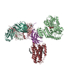 7rxcMC 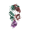 7r9dC 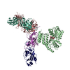 7rxdC M: atomic model generated by this map C: citing same article ( |
|---|---|
| Similar structure data |


-Links
| EMDB pages | EMDB (EBI/PDBe) / EMDataResource |
|---|---|
| Related items in Molecule of the Month |


-Map
| File | Download / File: emd_24728.map.gz / Format: CCP4 / Size: 46.4 MB / Type: IMAGE STORED AS FLOATING POINT NUMBER (4 BYTES) | ||||||||||||||||||||||||||||||||||||||||||||||||||||||||||||||||||||
|---|---|---|---|---|---|---|---|---|---|---|---|---|---|---|---|---|---|---|---|---|---|---|---|---|---|---|---|---|---|---|---|---|---|---|---|---|---|---|---|---|---|---|---|---|---|---|---|---|---|---|---|---|---|---|---|---|---|---|---|---|---|---|---|---|---|---|---|---|---|
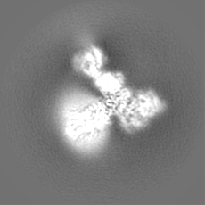
| Annotation | sharpened map for the complex | ||||||||||||||||||||||||||||||||||||||||||||||||||||||||||||||||||||
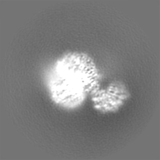
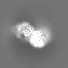
| Projections & slices | Image control
Images are generated by Spider. | ||||||||||||||||||||||||||||||||||||||||||||||||||||||||||||||||||||
| Voxel size | X=Y=Z: 1.06 Å | ||||||||||||||||||||||||||||||||||||||||||||||||||||||||||||||||||||
| Density |
| ||||||||||||||||||||||||||||||||||||||||||||||||||||||||||||||||||||
| Symmetry | Space group: 1 | ||||||||||||||||||||||||||||||||||||||||||||||||||||||||||||||||||||
| Details | EMDB XML:
CCP4 map header:
| ||||||||||||||||||||||||||||||||||||||||||||||||||||||||||||||||||||
 Z (Sec.)
 Y (Row.)
 X (Col.)


-Supplemental data
-Additional map: unsharpened map
| File | emd_24728_additional_1.map | ||||||||||||
|---|---|---|---|---|---|---|---|---|---|---|---|---|---|
| Annotation | unsharpened map | ||||||||||||
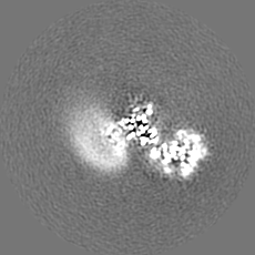
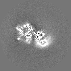
| Projections & Slices |
| ||||||||||||
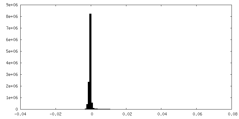
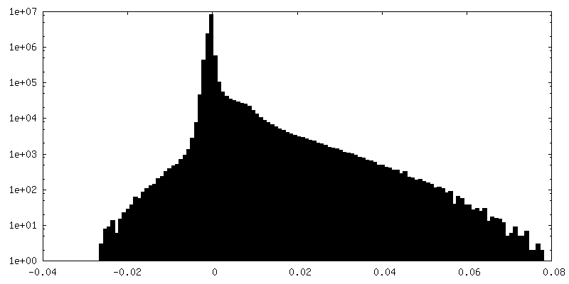
| Density Histograms |

- Sample components
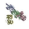
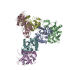
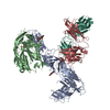
-Entire : The complex of KDELR with Legobody
| Entire | Name: The complex of KDELR with Legobody |
|---|---|
| Components |
|
-Supramolecule #1: The complex of KDELR with Legobody
| Supramolecule | Name: The complex of KDELR with Legobody / type: complex / ID: 1 / Parent: 0 / Macromolecule list: #1-#5 |
|---|---|
| Molecular weight | Theoretical: 150 KDa |
-Macromolecule #1: Fab_8D3_2 heavy chain
| Macromolecule | Name: Fab_8D3_2 heavy chain / type: protein_or_peptide / ID: 1 / Number of copies: 1 / Enantiomer: LEVO |
|---|---|
| Source (natural) | Organism: |
| Molecular weight | Theoretical: 25.252217 KDa |
| Recombinant expression | Organism:  Homo sapiens (human) Homo sapiens (human) |
| Sequence | String: DVQLVESGGG LVQPGKSLRL SCAASGFTFS NFGMHWVRQA PEMGLEWVAY ISSGSTTKYY GDTVKGRFTI SRDNPKNTLY LQMNSLRSE DTAMYYCARR PLYDGDYGYP MDYWGQGTSV TVSSASTKGP SVFPLAPSSK STSGGTAALG CLVKDYFPEP V TVSWNSGA ...String: DVQLVESGGG LVQPGKSLRL SCAASGFTFS NFGMHWVRQA PEMGLEWVAY ISSGSTTKYY GDTVKGRFTI SRDNPKNTLY LQMNSLRSE DTAMYYCARR PLYDGDYGYP MDYWGQGTSV TVSSASTKGP SVFPLAPSSK STSGGTAALG CLVKDYFPEP V TVSWNSGA LTSGVHTFPA VLQSSGLYSL SSVVTVPSSS LGTQTYICNV NHKPSNTKVD KKVEPKSCGS HHHHHH |
-Macromolecule #2: Fab_8D3_2 light chain
| Macromolecule | Name: Fab_8D3_2 light chain / type: protein_or_peptide / ID: 2 / Number of copies: 1 / Enantiomer: LEVO |
|---|---|
| Source (natural) | Organism: |
| Molecular weight | Theoretical: 24.095852 KDa |
| Recombinant expression | Organism: Homo sapiens (human) |
| Sequence | String: NIMLTQSPSS LAVSAGERVT MSCKSTQSIL YNSNQKTYLA WYQQKPGQSP KLLIYWASTR ASGVPDRFTG SGSGTDFTLT INSVQPEDL AVYYCHQYLS AWTFGGGTKL EIKRTVAAPS VFIFPPSDEQ LKSGTASVVC LLNNFYPREA KVQWKVDNAL Q SGNSQESV ...String: NIMLTQSPSS LAVSAGERVT MSCKSTQSIL YNSNQKTYLA WYQQKPGQSP KLLIYWASTR ASGVPDRFTG SGSGTDFTLT INSVQPEDL AVYYCHQYLS AWTFGGGTKL EIKRTVAAPS VFIFPPSDEQ LKSGTASVVC LLNNFYPREA KVQWKVDNAL Q SGNSQESV TEQDSKDSTY SLSSTLTLSK ADYEKHKVYA CEVTHQGLSS PVTKSFNRGE C |
-Macromolecule #3: Nb_KR
| Macromolecule | Name: Nb_KR / type: protein_or_peptide / ID: 3 / Number of copies: 1 / Enantiomer: LEVO |
|---|---|
| Source (natural) | Organism: |
| Molecular weight | Theoretical: 14.69826 KDa |
| Recombinant expression | Organism: |
| Sequence | String: QVQLVESGGG LVQAGGSLRL SCAASGFPVK RWSMTWYRQA PGKEREWVAA IRSAGHWTHY ADSVKGRFTI SRDNAKNTVY LQMNSLKPE DTAVYYCNVK DEGDFSYWYD YWGQGTQVTV SSLEHHHHHH |
-Macromolecule #4: Maltodextrin-binding protein,Immunoglobulin G-binding protein A,I...
| Macromolecule | Name: Maltodextrin-binding protein,Immunoglobulin G-binding protein A,Immunoglobulin G-binding protein G type: protein_or_peptide / ID: 4 / Number of copies: 1 / Enantiomer: LEVO |
|---|---|
| Source (natural) | Organism: Streptococcus sp. (bacteria) |
| Molecular weight | Theoretical: 59.233246 KDa |
| Recombinant expression | Organism: |
| Sequence | String: MKIEEGKLVI WINGDKGYNG LAEVGKKFEK DTGIKVTVEH PDKLEEKFPQ VAATGDGPDI IFWAHDRFGG YAQSGLLAEI TPDKAFQDK LYPFTWDAVR YNGKLIAYPI AVEALSLIYN KDLLPNPPKT WEEIPALDKE LKAKGKSALM FNLQEPYFTW P LIAADGGY ...String: MKIEEGKLVI WINGDKGYNG LAEVGKKFEK DTGIKVTVEH PDKLEEKFPQ VAATGDGPDI IFWAHDRFGG YAQSGLLAEI TPDKAFQDK LYPFTWDAVR YNGKLIAYPI AVEALSLIYN KDLLPNPPKT WEEIPALDKE LKAKGKSALM FNLQEPYFTW P LIAADGGY AFKYENGKYD IKDVGVDNAG AKAGLTFLVD LIKNKHMNAD TDYSIAEAAF NKGETAMTIN GPWAWSNIDT SK VNYGVTV LPTFKGQPSK PFVGVLSAGI NAASPNKELA KEFLENYLLT DEGLEAVNKD KPLGAVALKS YEEELAKDPR IAA TMENAQ KGEIMPNIPQ MSAFWYAVRT AVINAASGRQ TVDQALAFAQ ILIMPNLTEE QRNGFIQSLK DDPSVSKEIL AEAK KLNEH QAPKGGSGGA GSGDQQSAFY EILNMPNLNE AQRNGFIQSL KDDPSQSTNV LGEAKKLNES QAGGGSGGGS GGSAV TTYK LVINGKTLKG ETTTKAVDAE TAEKAFKQYA NDNGVDGVWT YDDATKTFTV TEGSGHHHHH H UniProtKB: Maltodextrin-binding protein, Immunoglobulin G-binding protein A, Immunoglobulin G-binding protein G |
-Macromolecule #5: ER lumen protein-retaining receptor 2
| Macromolecule | Name: ER lumen protein-retaining receptor 2 / type: protein_or_peptide / ID: 5 / Number of copies: 1 / Enantiomer: LEVO |
|---|---|
| Source (natural) | Organism: |
| Molecular weight | Theoretical: 30.083959 KDa |
| Recombinant expression | Organism:  |
| Sequence | String: MNIFRLTGDL SHLAAIIILL LKIWKSRSCA GISGKSQLLF ALVFTTRYLD LFTSFISLYN TSMKLIYIAC SYATVYLIYM KFKATYDGN HDTFRVEFLI VPVGGLSFLV NHDFSPLEIL WTFSIYLESV AILPQLFMIS KTGEAETITT HYLFFLGLYR A LYLVNWIW ...String: MNIFRLTGDL SHLAAIIILL LKIWKSRSCA GISGKSQLLF ALVFTTRYLD LFTSFISLYN TSMKLIYIAC SYATVYLIYM KFKATYDGN HDTFRVEFLI VPVGGLSFLV NHDFSPLEIL WTFSIYLESV AILPQLFMIS KTGEAETITT HYLFFLGLYR A LYLVNWIW RYYFEGFFDL IAVVAGVVQT VLYCDFFYLY VTKVLKGKKL SLPAGSGGEN LYFQSGGGMD EKTTGWRGGH VV EGLAGEL EQLRARLEHH PQGQREP UniProtKB: ER lumen protein-retaining receptor 2 |
-Macromolecule #7: (2S)-3-(hexadecanoyloxy)-2-[(9Z)-octadec-9-enoyloxy]propyl 2-(tri...
| Macromolecule | Name: (2S)-3-(hexadecanoyloxy)-2-[(9Z)-octadec-9-enoyloxy]propyl 2-(trimethylammonio)ethyl phosphate type: ligand / ID: 7 / Number of copies: 4 / Formula: POV |
|---|---|
| Molecular weight | Theoretical: 760.076 Da |
| Chemical component information |  ChemComp-POV: |
-Experimental details
-Structure determination
| Method | cryo EM |
|---|---|
 Processing Processing | single particle reconstruction |
| Aggregation state | particle |
-Sample preparation
| Buffer | pH: 7.4 |
|---|---|
| Vitrification | Cryogen name: ETHANE |
- Electron microscopy
| Microscope | FEI TITAN KRIOS |
|---|---|
| Image recording | Film or detector model: GATAN K3 (6k x 4k) / Average electron dose: 51.44 e/Å2 |
| Electron beam | Acceleration voltage: 300 kV / Electron source:  FIELD EMISSION GUN FIELD EMISSION GUN |
| Electron optics | Illumination mode: FLOOD BEAM / Imaging mode: BRIGHT FIELD |
| Experimental equipment |  Model: Titan Krios / Image courtesy: FEI Company |
