 ムービー
ムービー コントローラー


+ データを開く

- 基本情報
基本情報
| 登録情報 | データベース: PDB / ID: 7k8m | |||||||||
|---|---|---|---|---|---|---|---|---|---|---|
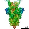
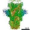
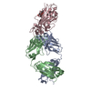
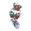
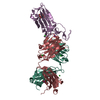
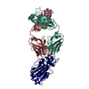
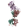
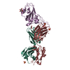
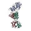
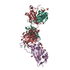
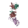
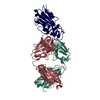
| タイトル | Structure of the SARS-CoV-2 receptor binding domain in complex with the human neutralizing antibody Fab fragment, C102 | |||||||||
 要素 要素 |
| |||||||||
 キーワード キーワード |  VIRAL PROTEIN/IMMUNE SYSTEM (ウイルス性) / Human Neutralizing Antibody (中和抗体) / SARS-CoV-2 (SARSコロナウイルス2) / Receptor Binding Domain (受容体) / COVID-19 (新型コロナウイルス感染症 (2019年)) / VIRAL PROTEIN-IMMUNE SYSTEM complex (ウイルス性) VIRAL PROTEIN/IMMUNE SYSTEM (ウイルス性) / Human Neutralizing Antibody (中和抗体) / SARS-CoV-2 (SARSコロナウイルス2) / Receptor Binding Domain (受容体) / COVID-19 (新型コロナウイルス感染症 (2019年)) / VIRAL PROTEIN-IMMUNE SYSTEM complex (ウイルス性) | |||||||||
| 機能・相同性 |  機能・相同性情報 機能・相同性情報Maturation of spike protein / viral translation / Translation of Structural Proteins / Virion Assembly and Release / host cell surface / host extracellular space / suppression by virus of host tetherin activity / Induction of Cell-Cell Fusion / structural constituent of virion / host cell endoplasmic reticulum-Golgi intermediate compartment membrane ...Maturation of spike protein / viral translation / Translation of Structural Proteins / Virion Assembly and Release / host cell surface / host extracellular space / suppression by virus of host tetherin activity / Induction of Cell-Cell Fusion / structural constituent of virion / host cell endoplasmic reticulum-Golgi intermediate compartment membrane / entry receptor-mediated virion attachment to host cell / receptor-mediated endocytosis of virus by host cell / Attachment and Entry / membrane fusion / positive regulation of viral entry into host cell / receptor-mediated virion attachment to host cell / receptor ligand activity / host cell surface receptor binding / fusion of virus membrane with host plasma membrane / fusion of virus membrane with host endosome membrane / エンベロープ (ウイルス) / symbiont-mediated suppression of host type I interferon-mediated signaling pathway / virion attachment to host cell / SARS-CoV-2 activates/modulates innate and adaptive immune responses / host cell plasma membrane / virion membrane / 生体膜 / identical protein binding / 細胞膜類似検索 - 分子機能 | |||||||||
| 生物種 |  Homo sapiens (ヒト) Homo sapiens (ヒト)  Severe acute respiratory syndrome coronavirus 2 (SARSコロナウイルス2) Severe acute respiratory syndrome coronavirus 2 (SARSコロナウイルス2) | |||||||||
| 手法 | X線回折 / シンクロトロン / 分子置換 / 解像度: 3.2 Å | |||||||||
 データ登録者 データ登録者 | Jette, C.A. / Barnes, C.O. / Bjorkman, P.J. | |||||||||
| 資金援助 |  米国, 2件 米国, 2件
| |||||||||
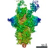
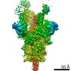
 引用 引用 | ジャーナル: Nature / 年: 2020 タイトル: SARS-CoV-2 neutralizing antibody structures inform therapeutic strategies. 著者: Christopher O Barnes / Claudia A Jette / Morgan E Abernathy / Kim-Marie A Dam / Shannon R Esswein / Harry B Gristick / Andrey G Malyutin / Naima G Sharaf / Kathryn E Huey-Tubman / Yu E Lee / ...著者: Christopher O Barnes / Claudia A Jette / Morgan E Abernathy / Kim-Marie A Dam / Shannon R Esswein / Harry B Gristick / Andrey G Malyutin / Naima G Sharaf / Kathryn E Huey-Tubman / Yu E Lee / Davide F Robbiani / Michel C Nussenzweig / Anthony P West / Pamela J Bjorkman /  要旨: The coronavirus disease 2019 (COVID-19) pandemic presents an urgent health crisis. Human neutralizing antibodies that target the host ACE2 receptor-binding domain (RBD) of the severe acute ...The coronavirus disease 2019 (COVID-19) pandemic presents an urgent health crisis. Human neutralizing antibodies that target the host ACE2 receptor-binding domain (RBD) of the severe acute respiratory syndrome coronavirus-2 (SARS-CoV-2) spike protein show promise therapeutically and are being evaluated clinically. Here, to identify the structural correlates of SARS-CoV-2 neutralization, we solved eight new structures of distinct COVID-19 human neutralizing antibodies in complex with the SARS-CoV-2 spike trimer or RBD. Structural comparisons allowed us to classify the antibodies into categories: (1) neutralizing antibodies encoded by the VH3-53 gene segment with short CDRH3 loops that block ACE2 and bind only to 'up' RBDs; (2) ACE2-blocking neutralizing antibodies that bind both up and 'down' RBDs and can contact adjacent RBDs; (3) neutralizing antibodies that bind outside the ACE2 site and recognize both up and down RBDs; and (4) previously described antibodies that do not block ACE2 and bind only to up RBDs. Class 2 contained four neutralizing antibodies with epitopes that bridged RBDs, including a VH3-53 antibody that used a long CDRH3 with a hydrophobic tip to bridge between adjacent down RBDs, thereby locking the spike into a closed conformation. Epitope and paratope mapping revealed few interactions with host-derived N-glycans and minor contributions of antibody somatic hypermutations to epitope contacts. Affinity measurements and mapping of naturally occurring and in vitro-selected spike mutants in 3D provided insight into the potential for SARS-CoV-2 to escape from antibodies elicited during infection or delivered therapeutically. These classifications and structural analyses provide rules for assigning current and future human RBD-targeting antibodies into classes, evaluating avidity effects and suggesting combinations for clinical use, and provide insight into immune responses against SARS-CoV-2. | |||||||||
| 履歴 |
|
- 構造の表示
構造の表示
| 構造ビューア | 分子: MolmilJmol/JSmol |
|---|
- ダウンロードとリンク
-ダウンロード
| PDBx/mmCIF形式 | 7k8m.cif.gz | 163.1 KB | 表示 | PDBx/mmCIF形式 |
|---|---|---|---|---|
| PDB形式 | pdb7k8m.ent.gz | 102.2 KB | 表示 | PDB形式 |
| PDBx/mmJSON形式 | 7k8m.json.gz | ツリー表示 | PDBx/mmJSON形式 | |
| その他 |  その他のダウンロード その他のダウンロード |
-検証レポート
| アーカイブディレクトリ | https://data.pdbj.org/pub/pdb/validation_reports/k8/7k8mftp://data.pdbj.org/pub/pdb/validation_reports/k8/7k8m | HTTPS FTP |
|---|
-関連構造データ
| 関連構造データ | 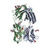 7k8nC 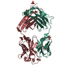 7k8oC 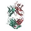 7k8pC 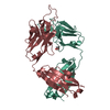 7k8qC 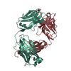 7k8rC  7k8sC  7k8tC 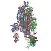 7k8uC  7k8vC  7k8wC  7k8xC  7k8yC  7k8zC  7k90C  7bz5S S: 精密化の開始モデル C: 同じ文献を引用 ( |
|---|---|
| 類似構造データ |

-リンク
 PDBj
PDBj


- 集合体
集合体
| 登録構造単位 | 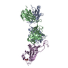
| ||||||||||||
|---|---|---|---|---|---|---|---|---|---|---|---|---|---|
| 1 |
| ||||||||||||
| 単位格子 |
|
-要素
| #1: 抗体 | 分子量: 24622.443 Da / 分子数: 1 / 由来タイプ: 組換発現 / 由来: (組換発現) Homo sapiens (ヒト) / 細胞株 (発現宿主): HEK293 / 発現宿主: Homo sapiens (ヒト) |
|---|---|
| #2: 抗体 | 分子量: 23417.938 Da / 分子数: 1 / 由来タイプ: 組換発現 / 由来: (組換発現) Homo sapiens (ヒト) / 細胞株 (発現宿主): HEK293 / 発現宿主: Homo sapiens (ヒト) |
| #3: タンパク質 | スパイクタンパク質 / S glycoprotein / E2 / Peplomer protein 分子量: 21152.633 Da / 分子数: 1 / Fragment: receptor binding domain (UNP residues 331-517) / 由来タイプ: 組換発現 由来: (組換発現) Severe acute respiratory syndrome coronavirus 2 (SARSコロナウイルス2)遺伝子: S, 2 / 細胞株 (発現宿主): HEK293 / 発現宿主: Homo sapiens (ヒト) / 参照: UniProt: P0DTC2 |
| #4: 糖 | ChemComp-NAG / N-アセチルグルコサミン  タイプ: D-saccharide, beta linking / 分子量: 221.208 Da / 分子数: 1 / 由来タイプ: 合成 / 式: C8H15NO6 タイプ: D-saccharide, beta linking / 分子量: 221.208 Da / 分子数: 1 / 由来タイプ: 合成 / 式: C8H15NO6 |
| 研究の焦点であるリガンドがあるか | N |
-実験情報
-実験
| 実験 | 手法: X線回折 / 使用した結晶の数: 1 |
|---|
- 試料調製
試料調製
| 結晶 | マシュー密度: 3.07 Å3/Da / 溶媒含有率: 59.98 % |
|---|---|
| 結晶化 | 温度: 298 K / 手法: 蒸気拡散法, シッティングドロップ法 / 詳細: 0.2 M sodium citrate tribasic, 20% w/v PEG3350 |
-データ収集
| 回折 | 平均測定温度: 100 K / Serial crystal experiment: N |
|---|---|
| 放射光源 | 由来: シンクロトロン / サイト: SSRL / ビームライン: BL12-1 / 波長: 0.9795 Å |
| 検出器 | タイプ: DECTRIS EIGER X 16M / 検出器: PIXEL / 日付: 2020年5月29日 |
| 放射 | プロトコル: SINGLE WAVELENGTH / 単色(M)・ラウエ(L): M / 散乱光タイプ: x-ray |
| 放射波長 | 波長: 0.9795 Å / 相対比: 1 |
| 反射 | 解像度: 3.2→52.12 Å / Num. obs: 14722 / % possible obs: 99.7 % / 冗長度: 6.5 % / Biso Wilson estimate: 77.79 Å2 / CC1/2: 0.987 / Rmerge(I) obs: 0.16 / Rpim(I) all: 0.068 / Net I/σ(I): 6 |
| 反射 シェル | 解像度: 3.2→3.31 Å / Rmerge(I) obs: 0.492 / Num. unique obs: 1413 / CC1/2: 0.974 / Rpim(I) all: 0.232 |
- 解析
解析
| ソフトウェア |
| ||||||||||||||||||||||||||||||||||||||||||||||||||||||||||||||||||||||||||||||||||||
|---|---|---|---|---|---|---|---|---|---|---|---|---|---|---|---|---|---|---|---|---|---|---|---|---|---|---|---|---|---|---|---|---|---|---|---|---|---|---|---|---|---|---|---|---|---|---|---|---|---|---|---|---|---|---|---|---|---|---|---|---|---|---|---|---|---|---|---|---|---|---|---|---|---|---|---|---|---|---|---|---|---|---|---|---|---|
| 精密化 | 構造決定の手法: 分子置換 開始モデル: PDB entry 7BZ5 解像度: 3.2→52.12 Å / SU ML: 0.3955 / 交差検証法: FREE R-VALUE / σ(F): 1.36 / 位相誤差: 22.0817 立体化学のターゲット値: GeoStd + Monomer Library + CDL v1.2
| ||||||||||||||||||||||||||||||||||||||||||||||||||||||||||||||||||||||||||||||||||||
| 溶媒の処理 | 減衰半径: 0.9 Å / VDWプローブ半径: 1.11 Å / 溶媒モデル: FLAT BULK SOLVENT MODEL | ||||||||||||||||||||||||||||||||||||||||||||||||||||||||||||||||||||||||||||||||||||
| 原子変位パラメータ | Biso mean: 73.34 Å2 | ||||||||||||||||||||||||||||||||||||||||||||||||||||||||||||||||||||||||||||||||||||
| 精密化ステップ | サイクル: LAST / 解像度: 3.2→52.12 Å
| ||||||||||||||||||||||||||||||||||||||||||||||||||||||||||||||||||||||||||||||||||||
| 拘束条件 |
| ||||||||||||||||||||||||||||||||||||||||||||||||||||||||||||||||||||||||||||||||||||
| LS精密化 シェル |
|
