| 登録情報 | データベース: PDB / ID: 6k1z
|
|---|
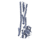
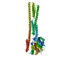
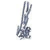
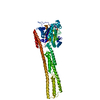
| タイトル | Crystal structure of farnesylated hGBP1 |
|---|
 要素 要素 | Guanylate-binding protein 1 |
|---|
 キーワード キーワード | HYDROLASE / GBP farnesylation |
|---|
| 機能・相同性 |  機能・相同性情報 機能・相同性情報
GDP phosphatase activity / non-canonical inflammasome complex assembly / protein localization to vacuole / negative regulation of substrate adhesion-dependent cell spreading / symbiont cell surface / cytolysis in another organism / positive regulation of pyroptotic inflammatory response / vesicle membrane / negative regulation of protein localization to plasma membrane / negative regulation of T cell receptor signaling pathway ...GDP phosphatase activity / non-canonical inflammasome complex assembly / protein localization to vacuole / negative regulation of substrate adhesion-dependent cell spreading / symbiont cell surface / cytolysis in another organism / positive regulation of pyroptotic inflammatory response / vesicle membrane / negative regulation of protein localization to plasma membrane / negative regulation of T cell receptor signaling pathway / spectrin binding / negative regulation of interleukin-2 production / defense response to protozoan / cytokine binding / 加水分解酵素; 酸無水物に作用; リン含有酸無水物に作用 / cellular response to interleukin-1 / regulation of protein localization to plasma membrane / regulation of calcium-mediated signaling / G protein activity / lipopolysaccharide binding / 加水分解酵素; 酸無水物に作用; GTPに作用・細胞または細胞小器官の運動に関与 / Hsp90 protein binding / cytoplasmic vesicle membrane / negative regulation of ERK1 and ERK2 cascade / cellular response to type II interferon / GDP binding / Interferon gamma signaling / actin cytoskeleton / cellular response to tumor necrosis factor / actin binding / cytoplasmic vesicle / defense response to virus / defense response to bacterium / Golgi membrane / innate immune response / GTPase activity / GTP binding / enzyme binding / Golgi apparatus / protein homodimerization activity / extracellular region / identical protein binding / plasma membrane / cytoplasm / cytosol類似検索 - 分子機能 Signaling Protein - Interferon-induced Guanylate-binding Protein 1; Chain A, domain 1 / Guanylate-binding protein, C-terminal domain / Guanylate-binding protein, C-terminal / Guanylate-binding protein/Atlastin, C-terminal / Guanylate-binding protein, C-terminal domain / Guanylate-binding protein, N-terminal / Guanylate-binding protein, C-terminal domain superfamily / Guanylate-binding protein, N-terminal domain / GB1/RHD3-type guanine nucleotide-binding (G) domain / GB1/RHD3-type guanine nucleotide-binding (G) domain profile. ...Signaling Protein - Interferon-induced Guanylate-binding Protein 1; Chain A, domain 1 / Guanylate-binding protein, C-terminal domain / Guanylate-binding protein, C-terminal / Guanylate-binding protein/Atlastin, C-terminal / Guanylate-binding protein, C-terminal domain / Guanylate-binding protein, N-terminal / Guanylate-binding protein, C-terminal domain superfamily / Guanylate-binding protein, N-terminal domain / GB1/RHD3-type guanine nucleotide-binding (G) domain / GB1/RHD3-type guanine nucleotide-binding (G) domain profile. / P-loop containing nucleotide triphosphate hydrolases / Up-down Bundle / Rossmann fold / P-loop containing nucleoside triphosphate hydrolase / 3-Layer(aba) Sandwich / Mainly Alpha / Alpha Beta類似検索 - ドメイン・相同性 |
|---|
| 生物種 |  Homo sapiens (ヒト) Homo sapiens (ヒト) |
|---|
| 手法 |  X線回折 / シンクロトロン / 分子置換 / 解像度: 2.307 Å X線回折 / シンクロトロン / 分子置換 / 解像度: 2.307 Å |
|---|
 データ登録者 データ登録者 | Du, S. / Xiao, J.Y. |
|---|
 引用 引用 | ジャーナル: Plos Pathog. / 年: 2019
タイトル: Structural mechanism for guanylate-binding proteins (GBPs) targeting by the Shigella E3 ligase IpaH9.8.
著者: Ji, C. / Du, S. / Li, P. / Zhu, Q. / Yang, X. / Long, C. / Yu, J. / Shao, F. / Xiao, J.Y. |
|---|
| 履歴 | | 登録 | 2019年5月13日 | 登録サイト: PDBJ / 処理サイト: PDBJ |
|---|
| 改定 1.0 | 2019年6月12日 | Provider: repository / タイプ: Initial release |
|---|
| 改定 1.1 | 2019年12月25日 | Group: Database references / カテゴリ: citation / citation_author
Item: _citation.country / _citation.journal_abbrev ..._citation.country / _citation.journal_abbrev / _citation.journal_id_CSD / _citation.journal_id_ISSN / _citation.journal_volume / _citation.page_first / _citation.page_last / _citation.pdbx_database_id_DOI / _citation.pdbx_database_id_PubMed / _citation.title / _citation.year |
|---|
|
|---|
 ムービー
ムービー コントローラー


 データを開く
 基本情報
基本情報 構造の表示
構造の表示 ダウンロードとリンク
 その他のダウンロード


 PDBj
PDBj 集合体
集合体

 分子量: 206.367 Da / 分子数: 1 / 由来タイプ: 合成 / 式: C15H26 / タイプ: SUBJECT OF INVESTIGATION
 分子量: 18.015 Da / 分子数: 128 / 由来タイプ: 天然 / 式: H2O
 試料調製
試料調製 / ビームライン: BL17U1 / 波長: 1 Å
 解析
解析