viral scaffold / protein localization to astral microtubule / regulation of slow-twitch skeletal muscle fiber contraction / cortical microtubule cytoskeleton / regulation of the force of skeletal muscle contraction / mitotic spindle astral microtubule end / protein localization to microtubule / muscle myosin complex / microtubule plus-end / muscle filament sliding ...viral scaffold / protein localization to astral microtubule / regulation of slow-twitch skeletal muscle fiber contraction / cortical microtubule cytoskeleton / regulation of the force of skeletal muscle contraction / mitotic spindle astral microtubule end / protein localization to microtubule / muscle myosin complex / microtubule plus-end / muscle filament sliding / cell projection membrane / regulation of the force of heart contraction / transition between fast and slow fiber / myosin filament / attachment of mitotic spindle microtubules to kinetochore / microtubule plus-end binding / myosin II complex / non-motile cilium assembly / adult heart development / microtubule bundle formation / cardiac muscle hypertrophy in response to stress / protein localization to centrosome / myosin complex / sarcomere organization / 微小管形成中心 / virion assembly / negative regulation of microtubule polymerization / microfilament motor activity / ventricular cardiac muscle tissue morphogenesis / myofibril / mitotic spindle pole / 微小管 / establishment of mitotic spindle orientation / skeletal muscle contraction / striated muscle contraction / spindle assembly / regulation of microtubule polymerization or depolymerization / spindle midzone / cytoplasmic microtubule / Amplification of signal from unattached kinetochores via a MAD2 inhibitory signal / stress fiber / ATP metabolic process / Mitotic Prometaphase / EML4 and NUDC in mitotic spindle formation / positive regulation of microtubule polymerization / cardiac muscle contraction / Loss of Nlp from mitotic centrosomes / Loss of proteins required for interphase microtubule organization from the centrosome / Recruitment of mitotic centrosome proteins and complexes / Resolution of Sister Chromatid Cohesion / Recruitment of NuMA to mitotic centrosomes / Anchoring of the basal body to the plasma membrane / regulation of heart rate / sarcomere / AURKA Activation by TPX2 / ciliary basal body / muscle contraction / RHO GTPases Activate Formins / protein localization / Z disc / Separation of Sister Chromatids / The role of GTSE1 in G2/M progression after G2 checkpoint / Regulation of PLK1 Activity at G2/M Transition / actin filament binding / 遊走 / 微小管 / molecular adaptor activity / calmodulin binding / cadherin binding / 細胞分裂 / focal adhesion / 中心体 / protein kinase binding / ゴルジ体 / DNA binding / RNA binding / ATP binding / identical protein binding / 細胞質基質 / 細胞質 類似検索 - 分子機能
Bacteriophage phi-29 scaffolding protein Gp7 / Capsid assembly scaffolding protein Gp7 / Phi29 scaffolding protein / EB1, C-terminal / Microtubule-associated protein RP/EB / EB1, C-terminal domain superfamily / EB1-C terminal (EB1-C) domain profile. / EB1-like C-terminal motif / DNA repair protein XRCC4-like, C-terminal / Myosin tail ...Bacteriophage phi-29 scaffolding protein Gp7 / Capsid assembly scaffolding protein Gp7 / Phi29 scaffolding protein / EB1, C-terminal / Microtubule-associated protein RP/EB / EB1, C-terminal domain superfamily / EB1-C terminal (EB1-C) domain profile. / EB1-like C-terminal motif / DNA repair protein XRCC4-like, C-terminal / Myosin tail / Myosin tail / Myosin N-terminal SH3-like domain / Myosin S1 fragment, N-terminal / Calponin homology (CH) domain / Myosin, N-terminal, SH3-like / Myosin N-terminal SH3-like domain profile. / Calponin homology domain / CH domain superfamily / Calponin homology (CH) domain profile. / Short calmodulin-binding motif containing conserved Ile and Gln residues. / Myosin head, motor domain / Myosin head (motor domain) / Myosin motor domain profile. / Myosin. Large ATPases. / IQ motif profile. / IQ motif, EF-hand binding site / Kinesin motor domain superfamily / P-loop containing nucleoside triphosphate hydrolase 類似検索 - ドメイン・相同性
Myosin-7 / Capsid assembly scaffolding protein / Microtubule-associated protein RP/EB family member 1 類似検索 - 構成要素
#241 - 2020年1月 20年の分子を振り返って (Twenty Years of Molecules) 類似性 (1)
-
集合体
登録構造単位
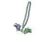
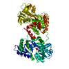
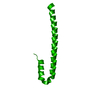
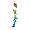
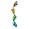
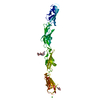
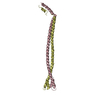
A: Gp7-MYH7(1173-1238)-EB1 chimera protein B: Gp7-MYH7(1173-1238)-EB1 chimera protein C: Gp7-MYH7(1173-1238)-EB1 chimera protein D: Gp7-MYH7(1173-1238)-EB1 chimera protein
Gp7-MYH7(1173-1238)-EB1chimeraprotein / chimera protein of Head morphogenesis protein / Myosin-7 and Microtubule-associated protein RP/EB ...chimera protein of Head morphogenesis protein / Myosin-7 and Microtubule-associated protein RP/EB family member 1
 ムービー
ムービー コントローラー


 データを開く
 基本情報
基本情報 要素
要素 キーワード
キーワード MOTOR PROTEIN (モータータンパク質) /
  機能・相同性情報


 データ登録者
 米国, 1件
  引用
引用 構造の表示
構造の表示 ダウンロードとリンク
 その他のダウンロード
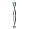


 PDBj
PDBj


 集合体
集合体


 分子量: 18.015 Da / 分子数: 35 / 由来タイプ: 天然 / 式: H2O
 試料調製
試料調製 解析
解析