 ムービー
ムービー コントローラー


+ データを開く

- 基本情報
基本情報
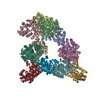
| 登録情報 | データベース: PDB / ID: 1i84 | ||||||
|---|---|---|---|---|---|---|---|
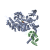
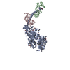
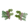
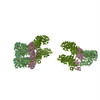
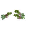
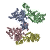
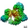
| タイトル | CRYO-EM STRUCTURE OF THE HEAVY MEROMYOSIN SUBFRAGMENT OF CHICKEN GIZZARD SMOOTH MUSCLE MYOSIN WITH REGULATORY LIGHT CHAIN IN THE DEPHOSPHORYLATED STATE. ONLY C ALPHAS PROVIDED FOR REGULATORY LIGHT CHAIN. ONLY BACKBONE ATOMS PROVIDED FOR S2 FRAGMENT. | ||||||
 要素 要素 |
| ||||||
 キーワード キーワード | CONTRACTILE PROTEIN / muscle protein / smooth muscle / myosin subfragment 2 / heavy meromyosin / essential light chain / regulatory light chain / motor protein / coiled-coil | ||||||
| 機能・相同性 |  機能・相同性情報 機能・相同性情報myosin II filament / myofibril assembly / elastic fiber assembly / myosin light chain binding / skeletal muscle myosin thick filament assembly / myosin II binding / muscle myosin complex / actomyosin / myosin filament / actomyosin structure organization ...myosin II filament / myofibril assembly / elastic fiber assembly / myosin light chain binding / skeletal muscle myosin thick filament assembly / myosin II binding / muscle myosin complex / actomyosin / myosin filament / actomyosin structure organization / myosin II complex / myosin complex / cardiac muscle cell development / structural constituent of muscle / microfilament motor activity / myofibril / myosin heavy chain binding / smooth muscle contraction / muscle contraction / skeletal muscle tissue development / ADP binding / actin filament binding / actin binding / calmodulin binding / calcium ion binding / magnesium ion binding / ATP binding / cytoplasm / cytosol 類似検索 - 分子機能 | ||||||
| 生物種 |  | ||||||
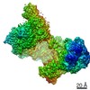
| 手法 | 電子線結晶学 / クライオ電子顕微鏡法 / 解像度: 20 Å | ||||||
 データ登録者 データ登録者 | Wendt, T. / Taylor, D. / Trybus, K.M. / Taylor, K. | ||||||
 引用 引用 | ジャーナル: Proc Natl Acad Sci U S A / 年: 2001 タイトル: Three-dimensional image reconstruction of dephosphorylated smooth muscle heavy meromyosin reveals asymmetry in the interaction between myosin heads and placement of subfragment 2. 著者: T Wendt / D Taylor / K M Trybus / K Taylor /  要旨: Regulation of the actin-activated ATPase of smooth muscle myosin II is known to involve an interaction between the two heads that is controlled by phosphorylation of the regulatory light chain. ...Regulation of the actin-activated ATPase of smooth muscle myosin II is known to involve an interaction between the two heads that is controlled by phosphorylation of the regulatory light chain. However, the three-dimensional structure of this inactivated form has been unknown. We have used a lipid monolayer to obtain two-dimensional crystalline arrays of the unphosphorylated inactive form of smooth muscle heavy meromyosin suitable for structural studies by electron cryomicroscopy of unstained, frozen-hydrated specimens. The three-dimensional structure reveals an asymmetric interaction between the two myosin heads. The ATPase activity of one head is sterically "blocked" because part of its actin-binding interface is positioned onto the converter domain of the second head. ATPase activity of the second head, which can bind actin, appears to be inhibited through stabilization of converter domain movements needed to release phosphate and achieve strong actin binding. When the subfragment 2 domain of heavy meromyosin is oriented as it would be in an actomyosin filament lattice, the position of the heads is very different from that needed to bind actin, suggesting an additional contribution to ATPase inhibition in situ. #1: ジャーナル: J.Cell Biol. / 年: 1999タイトル: Visualization of Head-head Interactions in the Inhibited State of Smooth Muscle Myosin 著者: Wendt, T. / Taylor, D. / Messier, T. / Trybus, K.M. / Taylor, K.A. #2: ジャーナル: Cell(Cambridge,Mass.) / 年: 1998タイトル: Crystal Structure of a Vertebrate Smooth Muscle Myosin Motor Domain and its Complex with the Essential Light Chain: Visualization of the Pre-power Stroke State 著者: Dominguez, R. / Freyzon, Y. / Trybus, K.M. / Cohen, C. #3: ジャーナル: Science / 年: 1993タイトル: Three-dimensional Structure of Myosin Subfragment-1: a Molecular Motor 著者: Rayment, I. / Rypniewsky, W.R. / Schmidt-Base, K. / Smith, R. / Tomchick, D.R. / Benning, M.M. / Winkelmann, D.A. / Wesenberg, G. / Holden, H.M. #4: ジャーナル: J.Mol.Biol. / 年: 1996タイトル: The Structure of the Head-tail Junction of the Myosin Molecule 著者: Offer, G. / Knight, P. | ||||||
| 履歴 |
|
- 構造の表示
構造の表示
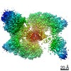
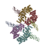
| ムービー |
ムービービューア |
|---|---|
| 構造ビューア | 分子: MolmilJmol/JSmol |
- ダウンロードとリンク
-ダウンロード
| PDBx/mmCIF形式 | 1i84.cif.gz | 400.3 KB | 表示 | PDBx/mmCIF形式 |
|---|---|---|---|---|
| PDB形式 | pdb1i84.ent.gz | 313.6 KB | 表示 | PDB形式 |
| PDBx/mmJSON形式 | 1i84.json.gz | ツリー表示 | PDBx/mmJSON形式 | |
| その他 |  その他のダウンロード その他のダウンロード |
-検証レポート
| 文書・要旨 | 1i84_validation.pdf.gz | 452.8 KB | 表示 | wwPDB検証レポート |
|---|---|---|---|---|
| 文書・詳細版 | 1i84_full_validation.pdf.gz | 678.8 KB | 表示 | |
| XML形式データ | 1i84_validation.xml.gz | 71.4 KB | 表示 | |
| CIF形式データ | 1i84_validation.cif.gz | 104.2 KB | 表示 | |
| アーカイブディレクトリ | https://data.pdbj.org/pub/pdb/validation_reports/i8/1i84ftp://data.pdbj.org/pub/pdb/validation_reports/i8/1i84 | HTTPS FTP |
-関連構造データ
| 関連構造データ | |
|---|---|
| 類似構造データ |

-リンク
 PDBj
PDBj


- 集合体
集合体
| 登録構造単位 | 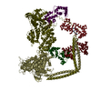
| ||||||||
|---|---|---|---|---|---|---|---|---|---|
| 1 |
| ||||||||
| 単位格子 |
|
-要素
| #1: 抗体 | 分子量: 137072.938 Da / 分子数: 2 / 断片: MEROMYOSIN SUBFRAGMENT. S1 AND S2 FRAGMENTS. / 由来タイプ: 組換発現 / 由来: (組換発現) 発現宿主:   Spodoptera frugiperda (ツマジロクサヨトウ) Spodoptera frugiperda (ツマジロクサヨトウ)株 (発現宿主): SF9 / 参照: UniProt: P10587, EC: 3.6.1.32 #2: タンパク質 | 分子量: 16873.025 Da / 分子数: 2 / 断片: S1 FRAGMENT / 由来タイプ: 組換発現 / 由来: (組換発現) 発現宿主: Spodoptera frugiperda (ツマジロクサヨトウ)株 (発現宿主): SF9 / 参照: GenBank: 212393, UniProt: P02607*PLUS #3: タンパク質 | 分子量: 18329.598 Da / 分子数: 2 / 断片: S1 FRAGMENT / 由来タイプ: 組換発現 / 由来: (組換発現) 発現宿主: Spodoptera frugiperda (ツマジロクサヨトウ)株 (発現宿主): SF9 / 参照: UniProt: P02609 |
|---|
-実験情報
-実験
| 実験 | 手法: 電子線結晶学 / 使用した結晶の数: 101 |
|---|---|
| EM実験 | 試料の集合状態: 2D ARRAY / 3次元再構成法: 電子線結晶学 |
| 結晶の対称性 | ∠γ: 91.5 ° / A: 133 Å / B: 304 Å / C: 90 Å / Space group name H-M: P2 |
- 試料調製
試料調製
| 構成要素 | 名称: HEAVY MEROMYOSIN SUBFRAGMENT OF CHICKEN GIZZARD SMOOTH MUSCLE MYOSIN WITH REGULATORY LIGHT CHAIN IN THE DEPHOSPHORYLATED STATE タイプ: COMPLEX |
|---|---|
| EM crystal formation | 詳細: 2-D arrays were grown on a positively charged lipid monolayer system consisting of didodecyldimethylammonium and dilauryl phosphatidyl choline (Taylor, K. A. & Taylor, D. W. (1992) J. Struct. ...詳細: 2-D arrays were grown on a positively charged lipid monolayer system consisting of didodecyldimethylammonium and dilauryl phosphatidyl choline (Taylor, K. A. & Taylor, D. W. (1992) J. Struct. Biol. 108, 140-147). Crystallization buffers consisted of 1 mM Mg2+, 20 mM phosphate, pH range 7-8, 1 mM ATP, 1 mM EGTA and PEG 6000 (7-10%) and NaCl (90-120 mM). Specimens were recovered on 200 mesh copper grids coated with a reticulated carbon film. The specimen was picked up from the grid bar side and blotted from that side. |
| 緩衝液 | pH: 7.5 |
| 試料 | 包埋: NO / シャドウイング: NO / 染色: NO / 凍結: YES |
| 急速凍結 | 装置: HOMEMADE PLUNGER / 凍結剤: ETHANE 詳細: Specimens were frozen in liquid ethane using a drop freezing apparatus. |
| 結晶化 | 温度: 277 K / 手法: lipid monolayer / pH: 7.5 詳細: MgCl, sodium phosphate, ATP, EGTA, PEG 6000, pH 7.5, Lipid monolayer, temperature 277K |
| 結晶化 | *PLUS 手法: other / 詳細: data none |
-データ収集
| 顕微鏡 | モデル: FEI/PHILIPS CM300FEG/T / 日付: 1999年7月1日 |
|---|---|
| 電子銃 | 加速電圧: 300 kV / 照射モード: FLOOD BEAM |
| 電子レンズ | モード: BRIGHT FIELD / 倍率(公称値): 24000 X |
| 試料ホルダ | 温度: 93 K |
| 撮影 | 電子線照射量: 5 e/Å2 / フィルム・検出器のモデル: KODAK SO-163 FILM |
| 画像スキャン | デジタル画像の数: 101 / Scanner model: PERKIN ELMER |
| 放射 | 散乱光タイプ: electron |
| 放射波長 | 相対比: 1 |
- 解析
解析
| 画像処理 | 詳細: Micrographs were processed using the ICE processing software | ||||||||||||
|---|---|---|---|---|---|---|---|---|---|---|---|---|---|
| 結晶の対称性 | ∠γ: 91.5 ° / A: 133 Å / B: 304 Å / C: 90 Å / Space group name H-M: P2 | ||||||||||||
| 3次元再構成 | 解像度: 20 Å / 解像度の算出法: DIFFRACTION PATTERN/LAYERLINES / 対称性のタイプ: 2D CRYSTAL | ||||||||||||
| 原子モデル構築 | プロトコル: OTHER / 空間: REAL / Target criteria: VISUAL AGREEMENT 詳細: REFINEMENT PROTOCOL--MANUAL DETAILS--The atomic model consists of two S1 heads and a segment of S2 containing 91 residues. Each S1 consists of a heavy chain and two light chains, the ELC and ...詳細: REFINEMENT PROTOCOL--MANUAL DETAILS--The atomic model consists of two S1 heads and a segment of S2 containing 91 residues. Each S1 consists of a heavy chain and two light chains, the ELC and the RLC. The atomic model of the S1 subfragment was constructed using two X-ray crystallography models. The heavy chain residues from ala2 to leu819 as well as the essential light chain were obtained from the chicken smooth muscle structure, PDB entry 1BR1, chains A and B. The remainder of S1 including heavy chain residues from glu820 to lys852 (smooth muscle numbering but skeletal myosin sequence) were obtained from residues glu811-lys843 of chicken skeletal muscle myosin, PDB entry 2MYS, since the structure of the entire smooth muscle S1 fragment is not available. Note that the residue numbers for the chain segment from 2MYS that were spliced onto 1BR1 have been renumbered to run consecutively from 819 but the residues themselves are the chicken skeletal muscle myosin heavy chain residues. The regulatory light chain coordinates were also obtained from 2MYS (skeletal) because a smooth muscle myosin model is not available. This regulatory light chain consisted only of C alphas. In fitting the model to the molecular envelope, the lever arm of the free head, i.e. ELC, RLC and heavy chain residues from ile796-lys852, were rotated as a rigid body about the Calpha of ile796. The amount of rotation was about 20 degrees. The blocked head was not modified. The alignment of 2MYS to 1BR1 was done using the conserved residues on the essential light chain. This alignment was carried out using O. The rms of the fit using 123 C alpha atoms of the conserved residues was 1.632 Angstroms. The C alpha distance between leu819 and glu811 (2MYS residue number) after the transformation was 4.23 Angstroms. This distance has not been altered in the model. The splice site at 819-820 is also slightly long and was left that way so the splice site would be easily visible. The other long bonds are at locations where the chain was bent to fit the structure. No minimization or refinement was done to fix them. The following residues have long peptide bond distances- chain S- LEU 819 and GLU 820 THR 854 and ARG 855 MET 860 and GLN 861 GLN 903 and ALA 904 chain V- LEU 819 and GLU 820 LYS 852 and VAL 853 MET 860 and GLN 861 GLN 903 and ALA 904 The 91 residues of the S2 fragment built into the model were calculated using a program written by Dr. Gerald Offer (reference 4). These coordinates consist only of backbone atoms. The S2 sequence begins at val853 which is the correct residue number for smooth muscle myosin. Peptide chain designations- Blocked myosin S1 head myosin heavy chain (with S2 segment) is chain V ELC is chain W RLC is chain Z Free myosin S1 head myosin heavy chain (with S2 fragment) is chain S ELC is chain T RLC is chain U The terms blocked and free refer to the conformations of the two S1 heads. SEQUENCE GAPS IN THE MOLECULAR MODEL- Myosin heavy chain- 205-210, 452-457, 635-655, 944-1185. ELC- 1-2. RLC (skeletal)- 1-18, 127, 142-147, 164-166. | ||||||||||||
| 精密化 | 最高解像度: 20 Å | ||||||||||||
| 精密化ステップ | サイクル: LAST / 最高解像度: 20 Å
|
