 Movie
Movie Controller
 Structure viewers
 About Yorodumi Papers


+Search query
-Structure paper
| Title | Three-dimensional image reconstruction of dephosphorylated smooth muscle heavy meromyosin reveals asymmetry in the interaction between myosin heads and placement of subfragment 2. |
|---|---|
| Journal, issue, pages | Proc Natl Acad Sci U S A, Vol. 98, Issue 8, Page 4361-4366, Year 2001 |
| Publish date | Apr 10, 2001 |
 Authors Authors | T Wendt / D Taylor / K M Trybus / K Taylor /  |
| PubMed Abstract | Regulation of the actin-activated ATPase of smooth muscle myosin II is known to involve an interaction between the two heads that is controlled by phosphorylation of the regulatory light chain. ...Regulation of the actin-activated ATPase of smooth muscle myosin II is known to involve an interaction between the two heads that is controlled by phosphorylation of the regulatory light chain. However, the three-dimensional structure of this inactivated form has been unknown. We have used a lipid monolayer to obtain two-dimensional crystalline arrays of the unphosphorylated inactive form of smooth muscle heavy meromyosin suitable for structural studies by electron cryomicroscopy of unstained, frozen-hydrated specimens. The three-dimensional structure reveals an asymmetric interaction between the two myosin heads. The ATPase activity of one head is sterically "blocked" because part of its actin-binding interface is positioned onto the converter domain of the second head. ATPase activity of the second head, which can bind actin, appears to be inhibited through stabilization of converter domain movements needed to release phosphate and achieve strong actin binding. When the subfragment 2 domain of heavy meromyosin is oriented as it would be in an actomyosin filament lattice, the position of the heads is very different from that needed to bind actin, suggesting an additional contribution to ATPase inhibition in situ. |
 External links External links | Proc Natl Acad Sci U S A / PubMed:11287639 / PubMed Central |
| Methods | EM (electron crystallography) |
| Resolution | 20 Å |
| Structure data | 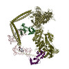 PDB-1i84: |
| Source |
|
 Keywords Keywords | CONTRACTILE PROTEIN / muscle protein / smooth muscle / myosin subfragment 2 / heavy meromyosin / essential light chain / regulatory light chain / motor protein / coiled-coil |

