ムービー
ムービー コントローラー


+ データを開く

- 基本情報
基本情報
| 登録情報 | データベース: EMDB / ID: EMD-5557 | |||||||||
|---|---|---|---|---|---|---|---|---|---|---|
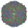
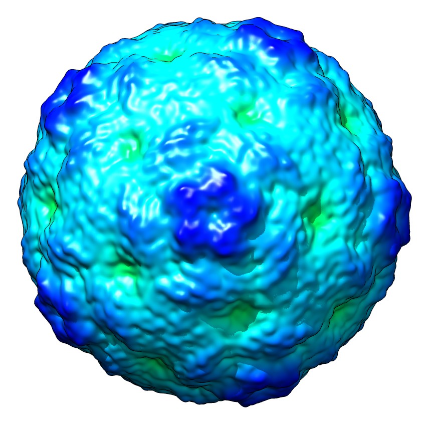
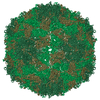
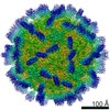
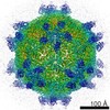
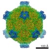
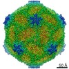
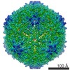
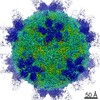
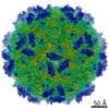
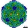
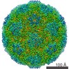
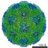
| タイトル | Structure of the EV71 strain 1095 procapsid | |||||||||
 マップデータ マップデータ | Reconstruction of EV71 strain 1095 procapsid | |||||||||
 試料 試料 |
| |||||||||
 キーワード キーワード |  picornavirus (ピコルナウイルス科) / enterovirus 71 / hand foot and mouth disease / cryo-EM (低温電子顕微鏡法) / single particle reconstruction (単粒子解析法) picornavirus (ピコルナウイルス科) / enterovirus 71 / hand foot and mouth disease / cryo-EM (低温電子顕微鏡法) / single particle reconstruction (単粒子解析法) | |||||||||
| 生物種 |   Human enterovirus 71 (エンテロウイルス) Human enterovirus 71 (エンテロウイルス) | |||||||||
| 手法 | 単粒子再構成法 / クライオ電子顕微鏡法 / 解像度: 8.78 Å | |||||||||
 データ登録者 データ登録者 | Cifuente JO / Lee H / Yoder JD / Shingler KL / Carnegie MS / Yoder JL / Ashley RE / Makhov AM / Conway JF / Hafenstein S | |||||||||
 引用 引用 | ジャーナル: J Virol / 年: 2013 タイトル: Structures of the procapsid and mature virion of enterovirus 71 strain 1095. 著者: Javier O Cifuente / Hyunwook Lee / Joshua D Yoder / Kristin L Shingler / Michael S Carnegie / Jennifer L Yoder / Robert E Ashley / Alexander M Makhov / James F Conway / Susan Hafenstein /  要旨: Enterovirus 71 (EV71) is an important emerging human pathogen with a global distribution and presents a disease pattern resembling poliomyelitis with seasonal epidemics that include cases of severe ...Enterovirus 71 (EV71) is an important emerging human pathogen with a global distribution and presents a disease pattern resembling poliomyelitis with seasonal epidemics that include cases of severe neurological complications, such as acute flaccid paralysis. EV71 is a member of the Picornaviridae family, which consists of icosahedral, nonenveloped, single-stranded RNA viruses. Here we report structures derived from X-ray crystallography and cryoelectron microscopy (cryo-EM) for the 1095 strain of EV71, including a putative precursor in virus assembly, the procapsid, and the mature virus capsid. The cryo-EM map of the procapsid provides new structural information on portions of the capsid proteins VP0 and VP1 that are disordered in the higher-resolution crystal structures. Our structures solved from virus particles in solution are largely in agreement with those from prior X-ray crystallographic studies; however, we observe small but significant structural differences for the 1095 procapsid compared to a structure solved in a previous study (X. Wang, W. Peng, J. Ren, Z. Hu, J. Xu, Z. Lou, X. Li, W. Yin, X. Shen, C. Porta, T. S. Walter, G. Evans, D. Axford, R. Owen, D. J. Rowlands, J. Wang, D. I. Stuart, E. E. Fry, and Z. Rao, Nat. Struct. Mol. Biol. 19:424-429, 2012) for a different strain of EV71. For both EV71 strains, the procapsid is significantly larger in diameter than the mature capsid, unlike in any other picornavirus. Nonetheless, our results demonstrate that picornavirus capsid expansion is possible without RNA encapsidation and that picornavirus assembly may involve an inward radial collapse of the procapsid to yield the native virion. | |||||||||
| 履歴 |
|
- 構造の表示
構造の表示
| ムービー |
 ムービービューア ムービービューア |
|---|---|
| 構造ビューア | EMマップ: SurfViewMolmilJmol/JSmol |
| 添付画像 |
- ダウンロードとリンク
-EMDBアーカイブ
| マップデータ | emd_5557.map.gz | 36.7 MB | EMDBマップデータ形式 | |
|---|---|---|---|---|
| ヘッダ (付随情報) | emd-5557-v30.xmlemd-5557.xml | 11.3 KB 11.3 KB | 表示 表示 | EMDBヘッダ |
| 画像 |  emd_5557_1.jpg emd_5557_1.jpg | 127.6 KB | ||
| アーカイブディレクトリ |  http://ftp.pdbj.org/pub/emdb/structures/EMD-5557ftp://ftp.pdbj.org/pub/emdb/structures/EMD-5557 http://ftp.pdbj.org/pub/emdb/structures/EMD-5557ftp://ftp.pdbj.org/pub/emdb/structures/EMD-5557 | HTTPS FTP |
-関連構造データ
-リンク
| EMDBのページ | EMDB (EBI/PDBe) / EMDataResource |
|---|
-マップ
| ファイル | ダウンロード / ファイル: emd_5557.map.gz / 形式: CCP4 / 大きさ: 154.3 MB / タイプ: IMAGE STORED AS FLOATING POINT NUMBER (4 BYTES) | ||||||||||||||||||||||||||||||||||||||||||||||||||||||||||||||||||||
|---|---|---|---|---|---|---|---|---|---|---|---|---|---|---|---|---|---|---|---|---|---|---|---|---|---|---|---|---|---|---|---|---|---|---|---|---|---|---|---|---|---|---|---|---|---|---|---|---|---|---|---|---|---|---|---|---|---|---|---|---|---|---|---|---|---|---|---|---|---|
| 注釈 | Reconstruction of EV71 strain 1095 procapsid | ||||||||||||||||||||||||||||||||||||||||||||||||||||||||||||||||||||
| ボクセルのサイズ | X=Y=Z: 1.2545 Å | ||||||||||||||||||||||||||||||||||||||||||||||||||||||||||||||||||||
| 密度 |
| ||||||||||||||||||||||||||||||||||||||||||||||||||||||||||||||||||||
| 対称性 | 空間群: 1 | ||||||||||||||||||||||||||||||||||||||||||||||||||||||||||||||||||||
| 詳細 | EMDB XML:
CCP4マップ ヘッダ情報:
| ||||||||||||||||||||||||||||||||||||||||||||||||||||||||||||||||||||
-添付データ
- 試料の構成要素
-全体 : EV71 strain 1095 procapsid
| 全体 | 名称: EV71 strain 1095 procapsid |
|---|---|
| 要素 |
|
-超分子 #1000: EV71 strain 1095 procapsid
| 超分子 | 名称: EV71 strain 1095 procapsid / タイプ: sample / ID: 1000 / Number unique components: 1 |
|---|
-超分子 #1: Human enterovirus 71
| 超分子 | 名称: Human enterovirus 71 / タイプ: virus / ID: 1 / NCBI-ID: 39054 / 生物種: Human enterovirus 71 / データベース: NCBI / ウイルスタイプ: VIRION / ウイルス・単離状態: STRAIN / ウイルス・エンベロープ: No / ウイルス・中空状態: Yes |
|---|---|
| 宿主 | 生物種:  Homo sapiens (ヒト) / 別称: VERTEBRATES Homo sapiens (ヒト) / 別称: VERTEBRATES |
| 分子量 | 理論値: 8 MDa |
| ウイルス殻 | Shell ID: 1 / 直径: 300 Å / T番号(三角分割数): 1 |
-実験情報
-構造解析
| 手法 | クライオ電子顕微鏡法 |
|---|---|
 解析 解析 | 単粒子再構成法 |
| 試料の集合状態 | particle |
-試料調製
| 緩衝液 | pH: 7.4 / 詳細: 10mM Tris-HCl, 200mM NaCl, 50mM MgCl2 |
|---|---|
| 凍結 | 凍結剤: ETHANE-PROPANE MIXTURE / チャンバー内温度: 120 K / 装置: FEI VITROBOT MARK I |
- 電子顕微鏡法
| 顕微鏡 | FEI TECNAI F20 |
|---|---|
| 電子線 | 加速電圧: 200 kV / 電子線源: FIELD EMISSION GUN |
| 電子光学系 | 倍率(補正後): 50400 / 照射モード: FLOOD BEAM / 撮影モード: BRIGHT FIELDBright-field microscopy / Cs: 2.0 mm / 最大 デフォーカス(公称値): 3.91 µm / 最小 デフォーカス(公称値): 0.79 µm / 倍率(公称値): 50000 |
| 特殊光学系 | エネルギーフィルター - 名称: FEI |
| 試料ステージ | 試料ホルダーモデル: GATAN LIQUID NITROGEN |
| 温度 | 最低: 83 K |
| アライメント法 | Legacy - 非点収差: Objective lens astigmatism was corrected at 100,000 times magnification |
| 日付 | 2011年8月15日 |
| 撮影 | カテゴリ: FILM / フィルム・検出器のモデル: KODAK SO-163 FILM デジタル化 - スキャナー: NIKON SUPER COOLSCAN 9000 デジタル化 - サンプリング間隔: 6.35 µm / 実像数: 20 |
| 実験機器 |  モデル: Tecnai F20 / 画像提供: FEI Company |
-画像解析
| CTF補正 | 詳細: Each particle |
|---|---|
| 最終 角度割当 | 詳細: AUTO3DEM |
| 最終 再構成 | アルゴリズム: OTHER / 解像度のタイプ: BY AUTHOR / 解像度: 8.78 Å / 解像度の算出法: FSC 0.5 CUT-OFF / ソフトウェア - 名称: Auto3dem, EMAN2 / 使用した粒子像数: 8805 |
| 詳細 | Semi-automatic particle selection was performed using e2boxer.py to obtain the particle coordinates, followed by particle boxing, linearization, normalization, and apodization of the images using Robem. Defocus and astigmatism values to perform contrast transfer function (CTF) correction were assessed using Robem for the extracted particles. The icosahedrally averaged reconstructions were initiated using a random model generated with setup_rmc and reached better than 10 A resolution estimated at a Fourier Shell Correlation, FSC=0.5. For the last step of refinement, the final maps were CTF corrected using a B factor of 300 A2. |
-原子モデル構築 1
| 初期モデル | PDB ID: Chain - #0 - Chain ID: A / Chain - #1 - Chain ID: B / Chain - #2 - Chain ID: C |
|---|---|
| ソフトウェア | 名称: VEDA |
| 詳細 | Protocol: Rigid body |
| 精密化 | 空間: RECIPROCAL / プロトコル: RIGID BODY FIT / 当てはまり具合の基準: R-factor |