 Movie
Movie Controller


+ Open data

- Basic information
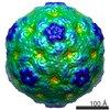
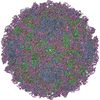
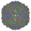
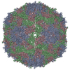
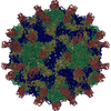
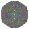
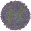
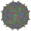
| Entry | Database: PDB / ID: 4gmp | ||||||
|---|---|---|---|---|---|---|---|
| Title | Crystal structure of enterovirus 71 strain 1095 procapsid | ||||||
 Components Components |
| ||||||
 Keywords Keywords | VIRUS / Capsid Protein | ||||||
| Function / homology |  Function and homology information Function and homology informationsymbiont-mediated suppression of host cytoplasmic pattern recognition receptor signaling pathway via inhibition of MDA-5 activity / picornain 2A / symbiont-mediated suppression of host mRNA export from nucleus / symbiont genome entry into host cell via pore formation in plasma membrane / picornain 3C / T=pseudo3 icosahedral viral capsid / host cell cytoplasmic vesicle membrane / virion component / ribonucleoside triphosphate phosphatase activity / host cell ...symbiont-mediated suppression of host cytoplasmic pattern recognition receptor signaling pathway via inhibition of MDA-5 activity / picornain 2A / symbiont-mediated suppression of host mRNA export from nucleus / symbiont genome entry into host cell via pore formation in plasma membrane / picornain 3C / T=pseudo3 icosahedral viral capsid / host cell cytoplasmic vesicle membrane / virion component / ribonucleoside triphosphate phosphatase activity / host cell / nucleoside-triphosphate phosphatase / channel activity / monoatomic ion transmembrane transport / DNA replication / RNA helicase activity / endocytosis involved in viral entry into host cell / symbiont-mediated activation of host autophagy / RNA-directed RNA polymerase / cysteine-type endopeptidase activity / viral RNA genome replication / RNA-directed RNA polymerase activity / symbiont entry into host cell / virion attachment to host cell / host cell nucleus / DNA-templated transcription / structural molecule activity / proteolysis / RNA binding / zinc ion binding / ATP binding Similarity search - Function | ||||||
| Biological species |   Human enterovirus 71 Human enterovirus 71 | ||||||
| Method |  X-RAY DIFFRACTION / SYNCHROTRON / MOLECULAR REPLACEMENT / Resolution: 3.9 Å X-RAY DIFFRACTION / SYNCHROTRON / MOLECULAR REPLACEMENT / Resolution: 3.9 Å | ||||||
 Authors Authors | Yoder, J.D. / Hafenstein, S. | ||||||
 Citation Citation | Journal: J Virol / Year: 2013 Title: Structures of the procapsid and mature virion of enterovirus 71 strain 1095. Authors: Javier O Cifuente / Hyunwook Lee / Joshua D Yoder / Kristin L Shingler / Michael S Carnegie / Jennifer L Yoder / Robert E Ashley / Alexander M Makhov / James F Conway / Susan Hafenstein /  Abstract: Enterovirus 71 (EV71) is an important emerging human pathogen with a global distribution and presents a disease pattern resembling poliomyelitis with seasonal epidemics that include cases of severe ...Enterovirus 71 (EV71) is an important emerging human pathogen with a global distribution and presents a disease pattern resembling poliomyelitis with seasonal epidemics that include cases of severe neurological complications, such as acute flaccid paralysis. EV71 is a member of the Picornaviridae family, which consists of icosahedral, nonenveloped, single-stranded RNA viruses. Here we report structures derived from X-ray crystallography and cryoelectron microscopy (cryo-EM) for the 1095 strain of EV71, including a putative precursor in virus assembly, the procapsid, and the mature virus capsid. The cryo-EM map of the procapsid provides new structural information on portions of the capsid proteins VP0 and VP1 that are disordered in the higher-resolution crystal structures. Our structures solved from virus particles in solution are largely in agreement with those from prior X-ray crystallographic studies; however, we observe small but significant structural differences for the 1095 procapsid compared to a structure solved in a previous study (X. Wang, W. Peng, J. Ren, Z. Hu, J. Xu, Z. Lou, X. Li, W. Yin, X. Shen, C. Porta, T. S. Walter, G. Evans, D. Axford, R. Owen, D. J. Rowlands, J. Wang, D. I. Stuart, E. E. Fry, and Z. Rao, Nat. Struct. Mol. Biol. 19:424-429, 2012) for a different strain of EV71. For both EV71 strains, the procapsid is significantly larger in diameter than the mature capsid, unlike in any other picornavirus. Nonetheless, our results demonstrate that picornavirus capsid expansion is possible without RNA encapsidation and that picornavirus assembly may involve an inward radial collapse of the procapsid to yield the native virion. | ||||||
| History |
|
- Structure visualization
| Structure viewer | Molecule: MolmilJmol/JSmol |
|---|
- Downloads & links
-Download
| PDBx/mmCIF format | 4gmp.cif.gz | 152.4 KB | Display | PDBx/mmCIF format |
|---|---|---|---|---|
| PDB format | pdb4gmp.ent.gz | 118.5 KB | Display | PDB format |
| PDBx/mmJSON format | 4gmp.json.gz | Tree view | PDBx/mmJSON format | |
| Others |  Other downloads Other downloads |
-Validation report
| Arichive directory | https://data.pdbj.org/pub/pdb/validation_reports/gm/4gmpftp://data.pdbj.org/pub/pdb/validation_reports/gm/4gmp | HTTPS FTP |
|---|
-Related structure data

-Links
 PDBj
PDBj

- Assembly
| Deposited unit | 
| ||||||||||||||||||||||||
|---|---|---|---|---|---|---|---|---|---|---|---|---|---|---|---|---|---|---|---|---|---|---|---|---|---|
| 1 | x 60
| ||||||||||||||||||||||||
| 2 |
| ||||||||||||||||||||||||
| 3 | x 5
| ||||||||||||||||||||||||
| 4 | x 6
| ||||||||||||||||||||||||
| 5 | 
| ||||||||||||||||||||||||
| 6 | x 5
| ||||||||||||||||||||||||
| Unit cell |
| ||||||||||||||||||||||||
| Symmetry | Point symmetry: (Schoenflies symbol: I (icosahedral)) | ||||||||||||||||||||||||
| Noncrystallographic symmetry (NCS) | NCS oper:
|
-Components
| #1: Protein | Mass: 35223.246 Da / Num. of mol.: 1 / Fragment: UNP residues 1-323 / Source method: isolated from a natural source / Source: (natural) Human enterovirus 71 / Strain: 1095 / References: UniProt: E5RPG0 |
|---|---|
| #2: Protein | Mass: 32646.758 Da / Num. of mol.: 1 / Fragment: UNP residues 566-862 / Source method: isolated from a natural source / Source: (natural) Human enterovirus 71 / Strain: 1095 / References: UniProt: E5RPG0, UniProt: Q91D09*PLUS |
| #3: Protein | Mass: 26466.227 Da / Num. of mol.: 1 / Fragment: UNP residues 324-565 / Source method: isolated from a natural source / Source: (natural) Human enterovirus 71 / Strain: 1095 / References: UniProt: E5RPG0 |
-Experimental details
-Experiment
| Experiment | Method: X-RAY DIFFRACTION / Number of used crystals: 1 |
|---|
- Sample preparation
| Crystal | Density Matthews: 3.8 Å3/Da / Density % sol: 67.59 % |
|---|---|
| Crystal grow | Temperature: 298 K / Method: vapor diffusion, hanging drop / pH: 7.5 Details: 0.4% PEG8000, 0.4% glycerol, 0.6 M sodium chloride, 0.2 M calcium chloride, 0.1 M Tris, pH 7.5, VAPOR DIFFUSION, HANGING DROP, temperature 298.0K |
-Data collection
| Diffraction | Mean temperature: 100 K |
|---|---|
| Diffraction source | Source: SYNCHROTRON / Site: CHESS / Beamline: F1 / Wavelength: 0.9179 Å |
| Detector | Type: ADSC QUANTUM 270 / Detector: CCD / Date: Jun 6, 2011 |
| Radiation | Monochromator: Horizontal bent Si(111), asymmetrically cut with water-cooled Cu block Protocol: SINGLE WAVELENGTH / Monochromatic (M) / Laue (L): M / Scattering type: x-ray |
| Radiation wavelength | Wavelength: 0.9179 Å / Relative weight: 1 |
| Reflection | Resolution: 3.9→50 Å / Num. obs: 66880 / % possible obs: 99.9 % / Observed criterion σ(I): -3 |
| Reflection shell | Resolution: 3.9→3.97 Å / % possible all: 100 |
- Processing
| Software |
| ||||||||||||||||||||||||
|---|---|---|---|---|---|---|---|---|---|---|---|---|---|---|---|---|---|---|---|---|---|---|---|---|---|
| Refinement | Method to determine structure: MOLECULAR REPLACEMENT / Resolution: 3.9→49.53 Å / Rfactor Rfree error: 0.005 / Data cutoff high absF: 18777118.35 / Data cutoff low absF: 0 / Isotropic thermal model: RESTRAINED / Cross valid method: THROUGHOUT / σ(F): 0 / Stereochemistry target values: Engh & Huber / Details: BULK SOLVENT MODEL USED
| ||||||||||||||||||||||||
| Solvent computation | Solvent model: FLAT MODEL / Bsol: 96.0406 Å2 / ksol: 0.34 e/Å3 | ||||||||||||||||||||||||
| Displacement parameters | Biso mean: 127.1 Å2
| ||||||||||||||||||||||||
| Refine analyze |
| ||||||||||||||||||||||||
| Refinement step | Cycle: LAST / Resolution: 3.9→49.53 Å
| ||||||||||||||||||||||||
| Refine LS restraints |
| ||||||||||||||||||||||||
| Refine LS restraints NCS | NCS model details: CONSTR | ||||||||||||||||||||||||
| LS refinement shell | Resolution: 3.9→4.14 Å / Rfactor Rfree error: 0.016 / Total num. of bins used: 6
| ||||||||||||||||||||||||
| Xplor file |
|
