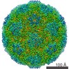
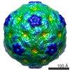
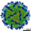
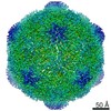
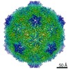
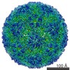
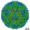
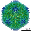
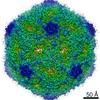
Journal: Nat Commun / Year: 2015 Title: Structure of Ljungan virus provides insight into genome packaging of this picornavirus. Authors: Ling Zhu / Xiangxi Wang / Jingshan Ren / Claudine Porta / Hannah Wenham / Jens-Ola Ekström / Anusha Panjwani / Nick J Knowles / Abhay Kotecha / C Alistair Siebert / A Michael Lindberg / ...Authors: Ling Zhu / Xiangxi Wang / Jingshan Ren / Claudine Porta / Hannah Wenham / Jens-Ola Ekström / Anusha Panjwani / Nick J Knowles / Abhay Kotecha / C Alistair Siebert / A Michael Lindberg / Elizabeth E Fry / Zihe Rao / Tobias J Tuthill / David I Stuart / Abstract: Picornaviruses are responsible for a range of human and animal diseases, but how their RNA genome is packaged remains poorly understood. A particularly poorly studied group within this family are ...Picornaviruses are responsible for a range of human and animal diseases, but how their RNA genome is packaged remains poorly understood. A particularly poorly studied group within this family are those that lack the internal coat protein, VP4. Here we report the atomic structure of one such virus, Ljungan virus, the type member of the genus Parechovirus B, which has been linked to diabetes and myocarditis in humans. The 3.78-Å resolution cryo-electron microscopy structure shows remarkable features, including an extended VP1 C terminus, forming a major protuberance on the outer surface of the virus, and a basic motif at the N terminus of VP3, binding to which orders some 12% of the viral genome. This apparently charge-driven RNA attachment suggests that this branch of the picornaviruses uses a different mechanism of genome encapsidation, perhaps explored early in the evolution of picornaviruses.
History
Deposition
Jul 21, 2015
-
Header (metadata) release
Sep 23, 2015
-
Map release
Oct 21, 2015
-
Update
Oct 21, 2015
-
Current status
Oct 21, 2015
Processing site: RCSB / Status: Released
-
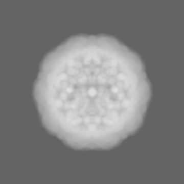
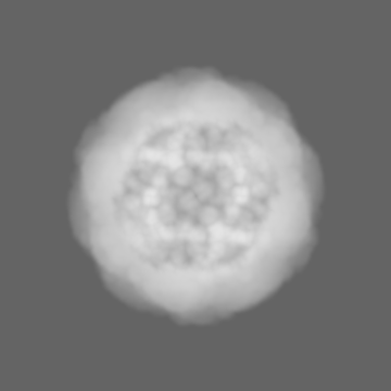
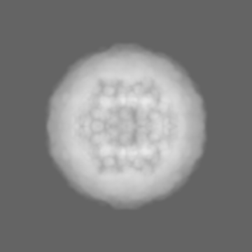
Structure visualization
Movie
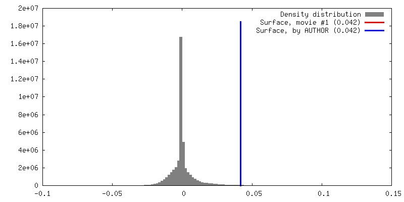
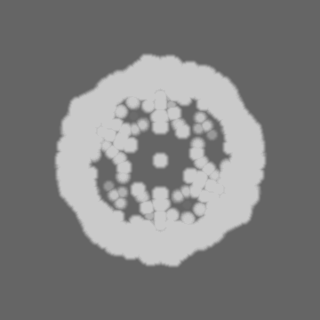
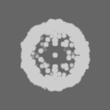
Surface view with section colored by density value
In the structure databanks used in Yorodumi, some data are registered as the other names, "COVID-19 virus" and "2019-nCoV". Here are the details of the virus and the list of structure data.
Jan 31, 2019. EMDB accession codes are about to change! (news from PDBe EMDB page)
EMDB accession codes are about to change! (news from PDBe EMDB page)
The allocation of 4 digits for EMDB accession codes will soon come to an end. Whilst these codes will remain in use, new EMDB accession codes will include an additional digit and will expand incrementally as the available range of codes is exhausted. The current 4-digit format prefixed with “EMD-” (i.e. EMD-XXXX) will advance to a 5-digit format (i.e. EMD-XXXXX), and so on. It is currently estimated that the 4-digit codes will be depleted around Spring 2019, at which point the 5-digit format will come into force.
The EM Navigator/Yorodumi systems omit the EMD- prefix.
Related info.:Q: What is EMD? / ID/Accession-code notation in Yorodumi/EM Navigator
Yorodumi is a browser for structure data from EMDB, PDB, SASBDB, etc.
This page is also the successor to EM Navigator detail page, and also detail information page/front-end page for Omokage search.
The word "yorodu" (or yorozu) is an old Japanese word meaning "ten thousand". "mi" (miru) is to see.
Related info.:EMDB / PDB / SASBDB / Comparison of 3 databanks / Yorodumi Search / Aug 31, 2016. New EM Navigator & Yorodumi / Yorodumi Papers / Jmol/JSmol / Function and homology information / Changes in new EM Navigator and Yorodumi
 Movie
Movie Controller


 Open data
 Basic information
 Map data
 Sample
 Keywords
 Function and homology information
 Ljungan virus
 Authors
 Citation


 Structure visualization
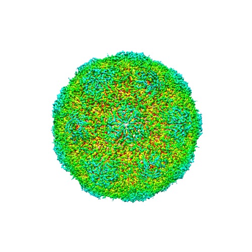
 Downloads & links
 http://ftp.pdbj.org/pub/emdb/structures/EMD-6394

 Z (Sec.)
 Y (Row.)
 X (Col.)


 Sample components
 Processing
 Electron microscopy
 FIELD EMISSION GUN
