 ムービー
ムービー コントローラー


+ データを開く

- 基本情報
基本情報
| 登録情報 | データベース: EMDB / ID: EMD-1605 | |||||||||
|---|---|---|---|---|---|---|---|---|---|---|
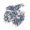
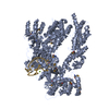
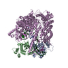
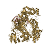
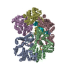
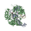
| タイトル | Solution structure of the KdpFABC P-type ATPase from Escherichia coli by electron microscopic single particle analysis | |||||||||
 マップデータ マップデータ | Volume of the KdpFABC P-type ATPase | |||||||||
 試料 試料 |
| |||||||||
 キーワード キーワード | KdpFABC /  P-type ATPase / potassium transport / single particle analysis (単粒子解析法) / electron microscopy. (電子顕微鏡) P-type ATPase / potassium transport / single particle analysis (単粒子解析法) / electron microscopy. (電子顕微鏡) | |||||||||
| 生物種 |  Escherichia coli (大腸菌) Escherichia coli (大腸菌) | |||||||||
| 手法 | 単粒子再構成法 / ネガティブ染色法 / 解像度: 19.0 Å | |||||||||
 データ登録者 データ登録者 | Heitkamp T / Bottcher B / Greie J-C | |||||||||
 引用 引用 | ジャーナル: J Struct Biol / 年: 2009 タイトル: Solution structure of the KdpFABC P-type ATPase from Escherichia coli by electron microscopic single particle analysis. 著者: Thomas Heitkamp / Bettina Böttcher / Jörg-Christian Greie /  要旨: The K+-translocating KdpFABC complex from Escherichia coli functions as a high affinity potassium uptake system and belongs to the superfamily of P-type ATPases, although it exhibits some unique ...The K+-translocating KdpFABC complex from Escherichia coli functions as a high affinity potassium uptake system and belongs to the superfamily of P-type ATPases, although it exhibits some unique features. It comprises four subunits, and the sites of ATP hydrolysis and substrate transport are located on two different polypeptides. No structural data are so far available for elucidating the correspondingly unique mechanism of coupling ion transport and catalysis in this P-type ATPase. By use of electron microscopy and single particle analysis of negatively stained, solubilized KdpFABC complexes, we solved the structure of the complex at a resolution of 19A, which allowed us to model the arrangement of subunits within the holoenzyme and, thus, to identify the interfaces between subunits. The model showed that the K+-translocating KdpA subunit is in close contact with the transmembrane region of the ATP-hydrolyzing subunit KdpB. The cytosolic C-terminal domain of the KdpC subunit, which is assumed to play a role in cooperative ATP binding together with KdpB, is located in close vicinity to the nucleotide binding domain of KdpB. Overall, the arrangement of subunits agrees with biochemical data and the predictions on subunit interactions. | |||||||||
| 履歴 |
|
- 構造の表示
構造の表示
| ムービー |
 ムービービューア ムービービューア |
|---|---|
| 構造ビューア | EMマップ: SurfViewMolmilJmol/JSmol |
| 添付画像 |
- ダウンロードとリンク
-EMDBアーカイブ
| マップデータ | emd_1605.map.gz | 167 KB | EMDBマップデータ形式 | |
|---|---|---|---|---|
| ヘッダ (付随情報) | emd-1605-v30.xmlemd-1605.xml | 11.9 KB 11.9 KB | 表示 表示 | EMDBヘッダ |
| 画像 |  image.gif image.gif | 91.4 KB | ||
| アーカイブディレクトリ |  http://ftp.pdbj.org/pub/emdb/structures/EMD-1605ftp://ftp.pdbj.org/pub/emdb/structures/EMD-1605 http://ftp.pdbj.org/pub/emdb/structures/EMD-1605ftp://ftp.pdbj.org/pub/emdb/structures/EMD-1605 | HTTPS FTP |
-関連構造データ


-リンク
| EMDBのページ | EMDB (EBI/PDBe) / EMDataResource |
|---|
-マップ
| ファイル | ダウンロード / ファイル: emd_1605.map.gz / 形式: CCP4 / 大きさ: 1.3 MB / タイプ: IMAGE STORED AS FLOATING POINT NUMBER (4 BYTES) | ||||||||||||||||||||||||||||||||||||||||||||||||||||||||||||||||||||
|---|---|---|---|---|---|---|---|---|---|---|---|---|---|---|---|---|---|---|---|---|---|---|---|---|---|---|---|---|---|---|---|---|---|---|---|---|---|---|---|---|---|---|---|---|---|---|---|---|---|---|---|---|---|---|---|---|---|---|---|---|---|---|---|---|---|---|---|---|---|
| 注釈 | Volume of the KdpFABC P-type ATPase | ||||||||||||||||||||||||||||||||||||||||||||||||||||||||||||||||||||
| 投影像・断面図 | 画像のコントロール
画像は Spider により作成 | ||||||||||||||||||||||||||||||||||||||||||||||||||||||||||||||||||||
| ボクセルのサイズ | X=Y=Z: 2.8 Å | ||||||||||||||||||||||||||||||||||||||||||||||||||||||||||||||||||||
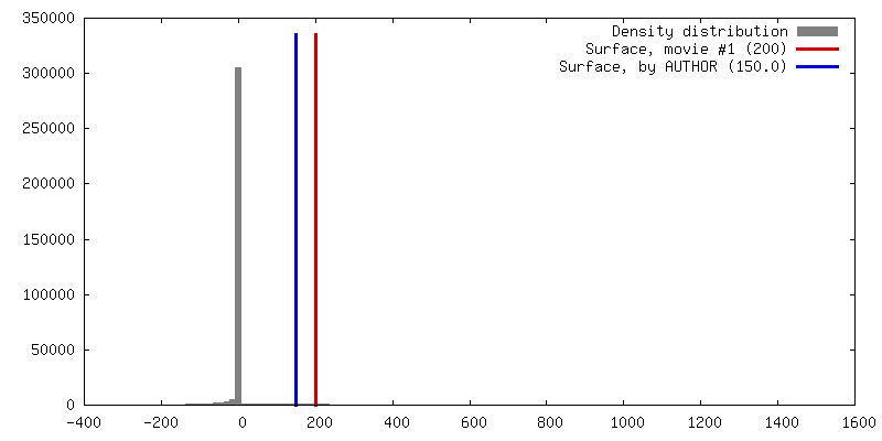
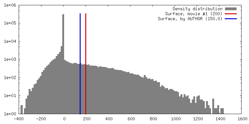
| 密度 |
| ||||||||||||||||||||||||||||||||||||||||||||||||||||||||||||||||||||
| 対称性 | 空間群: 1 | ||||||||||||||||||||||||||||||||||||||||||||||||||||||||||||||||||||
| 詳細 | EMDB XML:
CCP4マップ ヘッダ情報:
| ||||||||||||||||||||||||||||||||||||||||||||||||||||||||||||||||||||
 Z (Sec.)
 Y (Row.)
 X (Col.)


-添付データ
- 試料の構成要素
-全体 : KdpFABC P-type ATPase
| 全体 | 名称: KdpFABC P-type ATPase |
|---|---|
| 要素 |
|
-超分子 #1000: KdpFABC P-type ATPase
| 超分子 | 名称: KdpFABC P-type ATPase / タイプ: sample / ID: 1000 / 集合状態: monomer / Number unique components: 1 |
|---|---|
| 分子量 | 理論値: 154 KDa |
-分子 #1: KdpA-subunit
| 分子 | 名称: KdpA-subunit / タイプ: protein_or_peptide / ID: 1 / Name.synonym: KdpA-subunit / コピー数: 1 / 集合状態: monomer / 組換発現: Yes |
|---|---|
| 由来(天然) | 生物種: Escherichia coli (大腸菌) / 細胞中の位置: membrane |
| 分子量 | 理論値: 59 KDa |
| 組換発現 | 生物種: Escherichia coli (大腸菌) / 組換プラスミド: pGS4 |
-分子 #2: KdpB-subunit
| 分子 | 名称: KdpB-subunit / タイプ: protein_or_peptide / ID: 2 / Name.synonym: KdpB-subunit / コピー数: 1 / 集合状態: monomer / 組換発現: Yes |
|---|---|
| 由来(天然) | 生物種: Escherichia coli (大腸菌) / 細胞中の位置: membrane |
| 分子量 | 理論値: 72 KDa |
| 組換発現 | 生物種: Escherichia coli (大腸菌) / 組換プラスミド: pGS4 |
-分子 #3: KdpC-subunit
| 分子 | 名称: KdpC-subunit / タイプ: protein_or_peptide / ID: 3 / Name.synonym: KdpC-subunit / コピー数: 1 / 集合状態: monomer / 組換発現: Yes |
|---|---|
| 由来(天然) | 生物種: Escherichia coli (大腸菌) / 細胞中の位置: membrane |
| 分子量 | 理論値: 21 KDa |
| 組換発現 | 生物種: Escherichia coli (大腸菌) / 組換プラスミド: pGS4 |
-分子 #4: KdpF-subunit
| 分子 | 名称: KdpF-subunit / タイプ: protein_or_peptide / ID: 4 / Name.synonym: KdpF-subunit / コピー数: 1 / 集合状態: monomer / 組換発現: Yes |
|---|---|
| 由来(天然) | 生物種: Escherichia coli (大腸菌) / 細胞中の位置: membrane |
| 分子量 | 理論値: 3 KDa |
| 組換発現 | 生物種: Escherichia coli (大腸菌) / 組換プラスミド: pGS4 |
-実験情報
-構造解析
| 手法 | ネガティブ染色法 |
|---|---|
 解析 解析 | 単粒子再構成法 |
| 試料の集合状態 | particle |
-試料調製
| 濃度 | 0.005 mg/mL |
|---|---|
| 緩衝液 | pH: 6 詳細: 50 mM MES pH 6.0, 50 mM NaCl, 5 mM MgCl2, 5 mM CaCl2 |
| 染色 | タイプ: NEGATIVE 詳細: applied to freshly glow-discharged carbon-coated copper grids (400 mesh). Staining with 2 % (w/v) uranylacetic acid |
| グリッド | 詳細: 400 mesh copper grid, carbon coated |
| 凍結 | 凍結剤: NONE / 装置: OTHER |
- 電子顕微鏡法
| 顕微鏡 | FEI/PHILIPS CM120T |
|---|---|
| 電子線 | 加速電圧: 100 kV / 電子線源: LAB6 |
| 電子光学系 | 倍率(補正後): 48000 / 照射モード: FLOOD BEAM / 撮影モード: BRIGHT FIELDBright-field microscopy / Cs: 2 mm / 最大 デフォーカス(公称値): 0.864 µm / 最小 デフォーカス(公称値): 0.432 µm / 倍率(公称値): 52000 |
| 試料ステージ | 試料ホルダー: room temperature holder / 試料ホルダーモデル: SIDE ENTRY, EUCENTRIC |
| 温度 | 最低: 95 K / 最高: 295 K / 平均: 295 K |
| アライメント法 | Legacy - 非点収差: manually at 200000 on carbon film |
| 日付 | 2004年7月20日 |
| 撮影 | カテゴリ: FILM / フィルム・検出器のモデル: KODAK SO-163 FILM / デジタル化 - スキャナー: ZEISS SCAI / デジタル化 - サンプリング間隔: 14 µm / 実像数: 90 / ビット/ピクセル: 8 |
| Tilt angle min | 0 |
| Tilt angle max | 0 |
-画像解析
| 最終 再構成 | 想定した対称性 - 点群: C1 (非対称) / アルゴリズム: OTHER / 解像度のタイプ: BY AUTHOR / 解像度: 19.0 Å / 解像度の算出法: FSC 0.5 CUT-OFF / ソフトウェア - 名称: IMAGIC Spider / 使用した粒子像数: 10040 |
|---|---|
| 詳細 | started with angular reconstitution in IMAGIC (25 class averages) followed by projection matching in Spider (global search in 10 degree steps followed by local search in 2 degree steps) |
