 ムービー
ムービー コントローラー


+ データを開く

- 基本情報
基本情報
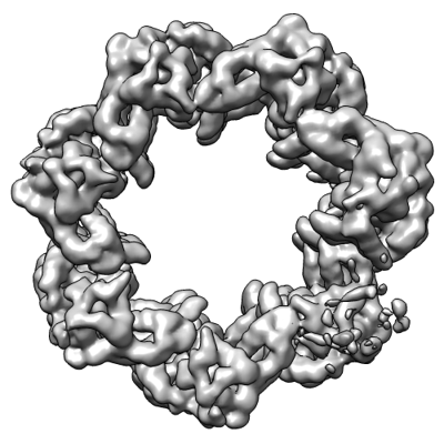
| 登録情報 | データベース: EMDB / ID: EMD-13355 | |||||||||
|---|---|---|---|---|---|---|---|---|---|---|
| タイトル | Structure of the Caulobacter crescentus S-layer protein RsaA N-terminal domain bound to LPS and soaked with Holmium | |||||||||
 マップデータ マップデータ | ||||||||||
 試料 試料 |
| |||||||||
 キーワード キーワード | S-layer protein RsaA bound to LPS and Holmium / STRUCTURAL PROTEIN | |||||||||
| 機能・相同性 | RsaA N-terminal domain / S-layer / RTX calcium-binding nonapeptide repeat / RTX calcium-binding nonapeptide repeat (4 copies) / Serralysin-like metalloprotease, C-terminal / calcium ion binding / extracellular region / S-layer protein 機能・相同性情報 機能・相同性情報 | |||||||||
| 生物種 |  Caulobacter vibrioides (バクテリア) Caulobacter vibrioides (バクテリア) | |||||||||
| 手法 | 単粒子再構成法 / クライオ電子顕微鏡法 / 解像度: 4.37 Å | |||||||||
 データ登録者 データ登録者 | von Kugelgen A / Bharat TAM | |||||||||
| 資金援助 |  英国, 2件 英国, 2件
| |||||||||
 引用 引用 | ジャーナル: Structure / 年: 2022 タイトル: High-resolution mapping of metal ions reveals principles of surface layer assembly in Caulobacter crescentus cells. 著者: Matthew Herdman / Andriko von Kügelgen / Danguole Kureisaite-Ciziene / Ramona Duman / Kamel El Omari / Elspeth F Garman / Andreas Kjaer / Dimitrios Kolokouris / Jan Löwe / Armin Wagner / ...著者: Matthew Herdman / Andriko von Kügelgen / Danguole Kureisaite-Ciziene / Ramona Duman / Kamel El Omari / Elspeth F Garman / Andreas Kjaer / Dimitrios Kolokouris / Jan Löwe / Armin Wagner / Phillip J Stansfeld / Tanmay A M Bharat / 要旨: Surface layers (S-layers) are proteinaceous crystalline coats that constitute the outermost component of most prokaryotic cell envelopes. In this study, we have investigated the role of metal ions in ...Surface layers (S-layers) are proteinaceous crystalline coats that constitute the outermost component of most prokaryotic cell envelopes. In this study, we have investigated the role of metal ions in the formation of the Caulobacter crescentus S-layer using high-resolution structural and cell biology techniques, as well as molecular simulations. Utilizing optical microscopy of fluorescently tagged S-layers, we show that calcium ions facilitate S-layer lattice formation and cell-surface binding. We report all-atom molecular dynamics simulations of the S-layer lattice, revealing the importance of bound metal ions. Finally, using electron cryomicroscopy and long-wavelength X-ray diffraction experiments, we mapped the positions of metal ions in the S-layer at near-atomic resolution, supporting our insights from the cellular and simulations data. Our findings contribute to the understanding of how C. crescentus cells form a regularly arranged S-layer on their surface, with implications on fundamental S-layer biology and the synthetic biology of self-assembling biomaterials. | |||||||||
| 履歴 |
|
- 構造の表示
構造の表示
| ムービー |
ムービービューア |
|---|---|
| 構造ビューア | EMマップ: SurfViewMolmilJmol/JSmol |
| 添付画像 |
- ダウンロードとリンク
-EMDBアーカイブ
| マップデータ | emd_13355.map.gz | 80.6 MB | EMDBマップデータ形式 | |
|---|---|---|---|---|
| ヘッダ (付随情報) | emd-13355-v30.xmlemd-13355.xml | 20.5 KB 20.5 KB | 表示 表示 | EMDBヘッダ |
| 画像 |  emd_13355.png emd_13355.png | 114.7 KB | ||
| Filedesc metadata | emd-13355.cif.gz | 7.7 KB | ||
| アーカイブディレクトリ |  http://ftp.pdbj.org/pub/emdb/structures/EMD-13355ftp://ftp.pdbj.org/pub/emdb/structures/EMD-13355 http://ftp.pdbj.org/pub/emdb/structures/EMD-13355ftp://ftp.pdbj.org/pub/emdb/structures/EMD-13355 | HTTPS FTP |
-検証レポート
| 文書・要旨 | emd_13355_validation.pdf.gz | 506.8 KB | 表示 | EMDB検証レポート |
|---|---|---|---|---|
| 文書・詳細版 | emd_13355_full_validation.pdf.gz | 506.4 KB | 表示 | |
| XML形式データ | emd_13355_validation.xml.gz | 6.3 KB | 表示 | |
| CIF形式データ | emd_13355_validation.cif.gz | 7.2 KB | 表示 | |
| アーカイブディレクトリ | https://ftp.pdbj.org/pub/emdb/validation_reports/EMD-13355ftp://ftp.pdbj.org/pub/emdb/validation_reports/EMD-13355 | HTTPS FTP |
-関連構造データ
| 関連構造データ |  7peoMC M: このマップから作成された原子モデル C: 同じ文献を引用 ( |
|---|---|
| 類似構造データ | |
| 電子顕微鏡画像生データ | EMPIAR-10790 (タイトル: High-resolution mapping of metal ions reveals principles of surface layer assembly in Caulobacter crescentus cells Data size: 304.4 Data #1: Unaligned multiframe micrographs of RsaA_NTD spiral soaked with 5 mM HoCl3 for 2 hours (converted from .mrc into .tif with relion_convert_to_tiff) [micrographs - multiframe] Data #2: Unaligned multiframe micrographs of RsaA_NTD spiral soaked with 5 mM HoCl3 for 2 hours with a 30 degree stage tilt (converted from .mrc into .tif with relion_convert_to_tiff) [micrographs - multiframe] Data #3: Aligned and dose-weighted micrographs of RsaA_NTD spiral soaked with 5 mM HoCl3 for 2 hours [micrographs - single frame] Data #4: Aligned and dose-weighted micrographs of RsaA_NTD spiral soaked with 5 mM HoCl3 for 2 hours with a 30 degree stage tilt [micrographs - single frame] Data #5: Gain reference image of the non-tilted and tilted dataset in MRC format [micrographs - single frame]) |


-リンク
| EMDBのページ | EMDB (EBI/PDBe) / EMDataResource |
|---|
-マップ
| ファイル | ダウンロード / ファイル: emd_13355.map.gz / 形式: CCP4 / 大きさ: 103 MB / タイプ: IMAGE STORED AS FLOATING POINT NUMBER (4 BYTES) | ||||||||||||||||||||||||||||||||||||||||||||||||||||||||||||||||||||
|---|---|---|---|---|---|---|---|---|---|---|---|---|---|---|---|---|---|---|---|---|---|---|---|---|---|---|---|---|---|---|---|---|---|---|---|---|---|---|---|---|---|---|---|---|---|---|---|---|---|---|---|---|---|---|---|---|---|---|---|---|---|---|---|---|---|---|---|---|---|
| ボクセルのサイズ | X=Y=Z: 1.08 Å | ||||||||||||||||||||||||||||||||||||||||||||||||||||||||||||||||||||
| 密度 |
| ||||||||||||||||||||||||||||||||||||||||||||||||||||||||||||||||||||
| 対称性 | 空間群: 1 | ||||||||||||||||||||||||||||||||||||||||||||||||||||||||||||||||||||
| 詳細 | EMDB XML:
CCP4マップ ヘッダ情報:
| ||||||||||||||||||||||||||||||||||||||||||||||||||||||||||||||||||||
-添付データ
- 試料の構成要素
-全体 : Structure of the Caulobacter crescentus S-layer protein RsaA N-te...
| 全体 | 名称: Structure of the Caulobacter crescentus S-layer protein RsaA N-terminal domain bound to LPS and soaked with Holmium |
|---|---|
| 要素 |
|
-超分子 #1: Structure of the Caulobacter crescentus S-layer protein RsaA N-te...
| 超分子 | 名称: Structure of the Caulobacter crescentus S-layer protein RsaA N-terminal domain bound to LPS and soaked with Holmium タイプ: complex / ID: 1 / 親要素: 0 / 含まれる分子: #1 詳細: Structure of the Caulobacter crescentus S-layer protein RsaA N-terminal domain bound to LPS and soaked with Holmium |
|---|---|
| 由来(天然) | 生物種: Caulobacter vibrioides (バクテリア) / 株: YB1001 |
-分子 #1: S-layer protein
| 分子 | 名称: S-layer protein / タイプ: protein_or_peptide / ID: 1 / 詳細: LPS O-antigen bound to the protein / コピー数: 1 / 光学異性体: LEVO |
|---|---|
| 由来(天然) | 生物種: Caulobacter vibrioides (バクテリア) / 株: YB1001 |
| 分子量 | 理論値: 25.820354 KDa |
| 組換発現 | 生物種: Caulobacter vibrioides CB15 (バクテリア) |
| 配列 | 文字列: AYTTAQLVTA YTNANLGKAP DAATTLTLDA YATQTQTGGL SDAAALTNTL KLVNSTTAVA IQTYQFFTGV APSAAGLDFL VDSTTNTND LNDAYYSKFA QENRFINFSI NLATGAGAGA TAFAAAYTGV SYAQTVATAY DKIIGNAVAT AAGVDVAAAV A FLSRQANI ...文字列: AYTTAQLVTA YTNANLGKAP DAATTLTLDA YATQTQTGGL SDAAALTNTL KLVNSTTAVA IQTYQFFTGV APSAAGLDFL VDSTTNTND LNDAYYSKFA QENRFINFSI NLATGAGAGA TAFAAAYTGV SYAQTVATAY DKIIGNAVAT AAGVDVAAAV A FLSRQANI DYLTAFVRAN TPFTAAADID LAVKAALIGT ILNAATVSGI GGYATATAAM INDLSDGALS TDNAAGVNLF TA YPSSGVS GSENLYFQ UniProtKB: S-layer protein |
-分子 #3: CALCIUM ION
| 分子 | 名称: CALCIUM ION / タイプ: ligand / ID: 3 / コピー数: 2 / 式: CA |
|---|---|
| 分子量 | 理論値: 40.078 Da |
-分子 #4: HOLMIUM ATOM
| 分子 | 名称: HOLMIUM ATOM / タイプ: ligand / ID: 4 / コピー数: 1 / 式: HO |
|---|---|
| 分子量 | 理論値: 164.93 Da |
| Chemical component information |  ChemComp-HO: |
-実験情報
-構造解析
| 手法 | クライオ電子顕微鏡法 |
|---|---|
 解析 解析 | 単粒子再構成法 |
| 試料の集合状態 | particle |
-試料調製
| 濃度 | 2.25 mg/mL | ||||||||||||||||||
|---|---|---|---|---|---|---|---|---|---|---|---|---|---|---|---|---|---|---|---|
| 緩衝液 | pH: 7.5 構成要素:
詳細: Buffer solutions were prepared fresh from sterile filtered concentrated stocksolutions. Solutions were filtered through a 0.22 um filter to avoid microbial contamination and degassed using a ...詳細: Buffer solutions were prepared fresh from sterile filtered concentrated stocksolutions. Solutions were filtered through a 0.22 um filter to avoid microbial contamination and degassed using a vacuum fold pump. The pH of the HEPES stock solution was adjusted with sodium hydroxide at 4 deg C. 5 mM HoCl3 was added to the specimen 1.5 hours before vitrification. | ||||||||||||||||||
| グリッド | モデル: Quantifoil R2/2 / 材質: COPPER/RHODIUM / メッシュ: 200 / 支持フィルム - 材質: CARBON / 支持フィルム - トポロジー: HOLEY ARRAY / 前処理 - タイプ: GLOW DISCHARGE / 前処理 - 時間: 20 sec. / 前処理 - 雰囲気: AIR / 詳細: 20 seconds, 15 mA | ||||||||||||||||||
| 凍結 | 凍結剤: ETHANE / チャンバー内湿度: 100 % / チャンバー内温度: 283.15 K / 装置: FEI VITROBOT MARK IV 詳細: Vitrobot options: Blot time 4 seconds, Blot force -13,1, Wait time 10 seconds, Drain time 0.5 seconds,. | ||||||||||||||||||
| 詳細 | RsaA N-terminal domain with LPS soaked with 5 mM HoCl3 for 1.5 h on ice before vitrification |
- 電子顕微鏡法
| 顕微鏡 | FEI TITAN KRIOS |
|---|---|
| 温度 | 最低: 70.0 K / 最高: 70.0 K |
| 特殊光学系 | エネルギーフィルター - 名称: GIF Quantum LS / エネルギーフィルター - スリット幅: 20 eV |
| 詳細 | EPU software |
| 撮影 | フィルム・検出器のモデル: GATAN K2 SUMMIT (4k x 4k) 検出モード: COUNTING / デジタル化 - サイズ - 横: 3838 pixel / デジタル化 - サイズ - 縦: 3710 pixel / デジタル化 - 画像ごとのフレーム数: 1-20 / 撮影したグリッド数: 2 / 実像数: 2038 / 平均露光時間: 8.0 sec. / 平均電子線量: 44.8 e/Å2 詳細: Two data collections: First: 0 degree stage tilt with 903 collected movies. Second: 30 degree stage tilt with 1135 collected movies |
| 電子線 | 加速電圧: 300 kV / 電子線源:  FIELD EMISSION GUN FIELD EMISSION GUN |
| 電子光学系 | C2レンズ絞り径: 50.0 µm / 最大 デフォーカス(補正後): -4.0 µm / 最小 デフォーカス(補正後): -1.0 µm / 倍率(補正後): 130000 / 照射モード: FLOOD BEAM / 撮影モード: BRIGHT FIELD / Cs: 2.7 mm / 最大 デフォーカス(公称値): -4.0 µm / 最小 デフォーカス(公称値): -1.0 µm / 倍率(公称値): 130000 |
| 試料ステージ | 試料ホルダーモデル: FEI TITAN KRIOS AUTOGRID HOLDER ホルダー冷却材: NITROGEN |
| 実験機器 |  モデル: Titan Krios / 画像提供: FEI Company |
+画像解析
-原子モデル構築 1
| 初期モデル | PDB ID: Chain - Chain ID: A / Chain - Source name: PDB / Chain - Initial model type: experimental model |
|---|---|
| 詳細 | The atomic coordinates (PDB ID 6T72) of our previous cryo-EM structure (von Kugelgen et al., 2020) of the RsaANTD oligomer bound to the O-antigen of lipopolysaccharide (LPS) were rigid body fitted into the final post-processed map from Relion 3.0 (Zivanov et al., 2018) using UCSF Chimera (Pettersen et al., 2004). The resulting fitted model was subjected to real-space refinement using Refmac5 (Murshudov et al., 2011) inside the CCP-EM suite (Burnely et al., 2017), as described previously (von Kugelgen et al., 2020), using reference restraints of the initial structure (PDB ID 6T72) generated with PROSMART (Nicholls et al. 2012). |
| 精密化 | 空間: REAL / プロトコル: RIGID BODY FIT |
| 得られたモデル | PDB-7peo: |

