 ムービー
ムービー コントローラー


+ データを開く

- 基本情報
基本情報
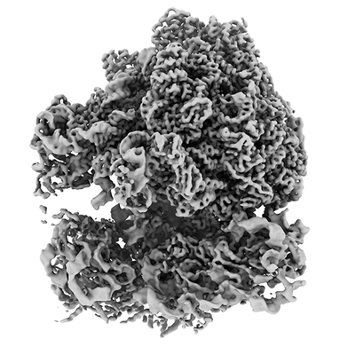
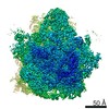
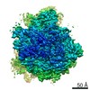
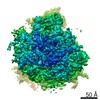
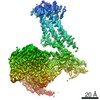
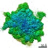
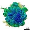
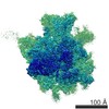
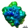
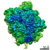
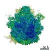
| 登録情報 | データベース: EMDB / ID: EMD-11999 | |||||||||||||||
|---|---|---|---|---|---|---|---|---|---|---|---|---|---|---|---|---|
| タイトル | M. pneumoniae 70S ribosome in complex with chloramphenicol obtained from in situ data using M, focused refinement of 50S sub-unit | |||||||||||||||
 マップデータ マップデータ | ||||||||||||||||
 試料 試料 |
| |||||||||||||||
| 機能・相同性 |  機能・相同性情報 機能・相同性情報large ribosomal subunit rRNA binding / large ribosomal subunit /  transferase activity / tRNA binding / rRNA binding / リボソーム / structural constituent of ribosome / 翻訳 (生物学) / ribonucleoprotein complex / response to antibiotic / 細胞質 transferase activity / tRNA binding / rRNA binding / リボソーム / structural constituent of ribosome / 翻訳 (生物学) / ribonucleoprotein complex / response to antibiotic / 細胞質類似検索 - 分子機能 | |||||||||||||||
| 生物種 |  Mycoplasma pneumoniae (バクテリア) Mycoplasma pneumoniae (バクテリア) | |||||||||||||||
| 手法 | サブトモグラム平均法 / クライオ電子顕微鏡法 / 解像度: 3.4 Å | |||||||||||||||
 データ登録者 データ登録者 | Tegunov D / Xue L / Dienemann C / Cramer P / Mahamid J | |||||||||||||||
| 資金援助 |  ドイツ, 4件 ドイツ, 4件
| |||||||||||||||
 引用 引用 | ジャーナル: Nat Methods / 年: 2021 タイトル: Multi-particle cryo-EM refinement with M visualizes ribosome-antibiotic complex at 3.5 Å in cells. 著者: Dimitry Tegunov / Liang Xue / Christian Dienemann / Patrick Cramer / Julia Mahamid / 要旨: Cryo-electron microscopy (cryo-EM) enables macromolecular structure determination in vitro and inside cells. In addition to aligning individual particles, accurate registration of sample motion and ...Cryo-electron microscopy (cryo-EM) enables macromolecular structure determination in vitro and inside cells. In addition to aligning individual particles, accurate registration of sample motion and three-dimensional deformation during exposures are crucial for achieving high-resolution reconstructions. Here we describe M, a software tool that establishes a reference-based, multi-particle refinement framework for cryo-EM data and couples a comprehensive spatial deformation model to in silico correction of electron-optical aberrations. M provides a unified optimization framework for both frame-series and tomographic tilt-series data. We show that tilt-series data can provide the same resolution as frame-series data on a purified protein specimen, indicating that the alignment step no longer limits the resolution obtainable from tomographic data. In combination with Warp and RELION, M resolves to residue level a 70S ribosome bound to an antibiotic inside intact bacterial cells. Our work provides a computational tool that facilitates structural biology in cells. | |||||||||||||||
| 履歴 |
|
- 構造の表示
構造の表示
| ムービー |
ムービービューア |
|---|---|
| 構造ビューア | EMマップ: SurfViewMolmilJmol/JSmol |
| 添付画像 |
- ダウンロードとリンク
-EMDBアーカイブ
| マップデータ | emd_11999.map.gz | 191.6 MB | EMDBマップデータ形式 | |
|---|---|---|---|---|
| ヘッダ (付随情報) | emd-11999-v30.xmlemd-11999.xml | 12.8 KB 12.8 KB | 表示 表示 | EMDBヘッダ |
| FSC (解像度算出) | emd_11999_fsc.xml | 13 KB | 表示 | FSCデータファイル |
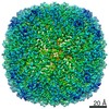
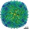
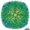
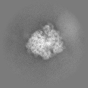
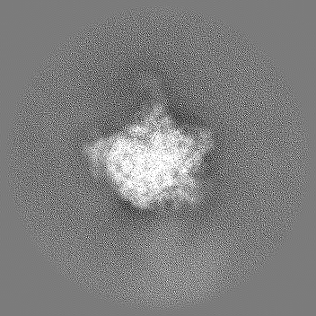
| 画像 |  emd_11999.png emd_11999.png | 85.2 KB | ||
| マスクデータ | emd_11999_msk_1.map | 166.4 MB | マスクマップ | |
| その他 | emd_11999_half_map_1.map.gzemd_11999_half_map_2.map.gz | 156.7 MB 156.7 MB | ||
| アーカイブディレクトリ |  http://ftp.pdbj.org/pub/emdb/structures/EMD-11999ftp://ftp.pdbj.org/pub/emdb/structures/EMD-11999 http://ftp.pdbj.org/pub/emdb/structures/EMD-11999ftp://ftp.pdbj.org/pub/emdb/structures/EMD-11999 | HTTPS FTP |
-関連構造データ
| 関連構造データ |  7oodM C: 同じ文献を引用 ( M: このマップから作成された原子モデル |
|---|---|
| 類似構造データ | |
| 電子顕微鏡画像生データ | EMPIAR-10499 (タイトル: Tilt series of native M. pneumoniae cells treated with chloramphenicol Data size: 83.8 Data #1: Unaligned tilt movies of M. pneumoniae [tilt series]) EMPIAR-10731 (タイトル: Locating Macromolecular Assemblies in Cells by 2D Template Matching with cisTEMData size: 67.8 Data #1: 50S templates of Mycoplasma pneumoniae used for 2D and 3D template matching [reconstructed volumes] Data #2: Single-exposure micrographs of untilted Mycoplasma pneumoniae cells [micrographs - single frame] Data #3: Tomographic tilt series of Mycoplasma pneumoniae cells [tilt series] Data #4: Tomographic reconstructions of Mycoplasma pneumoniae cells [reconstructed volumes]) |


-リンク
| EMDBのページ | EMDB (EBI/PDBe) / EMDataResource |
|---|---|
| 「今月の分子」の関連する項目 |


-マップ
| ファイル | ダウンロード / ファイル: emd_11999.map.gz / 形式: CCP4 / 大きさ: 209.3 MB / タイプ: IMAGE STORED AS FLOATING POINT NUMBER (4 BYTES) | ||||||||||||||||||||||||||||||||||||||||||||||||||||||||||||||||||||
|---|---|---|---|---|---|---|---|---|---|---|---|---|---|---|---|---|---|---|---|---|---|---|---|---|---|---|---|---|---|---|---|---|---|---|---|---|---|---|---|---|---|---|---|---|---|---|---|---|---|---|---|---|---|---|---|---|---|---|---|---|---|---|---|---|---|---|---|---|---|
| ボクセルのサイズ | X=Y=Z: 0.8502 Å | ||||||||||||||||||||||||||||||||||||||||||||||||||||||||||||||||||||
| 密度 |
| ||||||||||||||||||||||||||||||||||||||||||||||||||||||||||||||||||||
| 対称性 | 空間群: 1 | ||||||||||||||||||||||||||||||||||||||||||||||||||||||||||||||||||||
| 詳細 | EMDB XML:
CCP4マップ ヘッダ情報:
| ||||||||||||||||||||||||||||||||||||||||||||||||||||||||||||||||||||
-添付データ
-マスク #1
| ファイル | emd_11999_msk_1.map | ||||||||||||
|---|---|---|---|---|---|---|---|---|---|---|---|---|---|
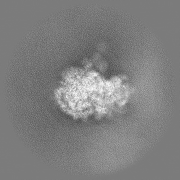
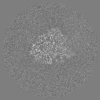
| 投影像・断面図 |
| ||||||||||||
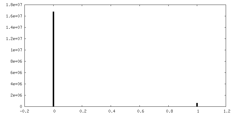
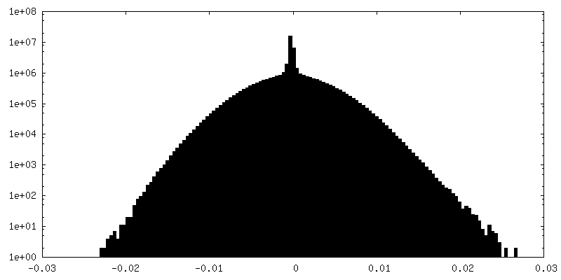
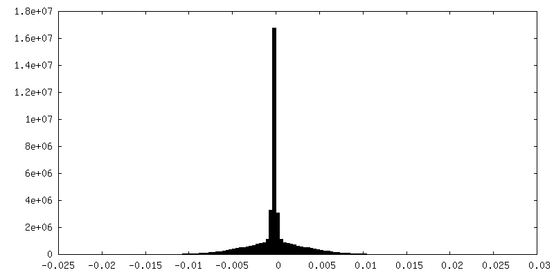
| 密度ヒストグラム |
 Z
Z Y
Y X
X


-ハーフマップ: #1
| ファイル | emd_11999_half_map_1.map | ||||||||||||
|---|---|---|---|---|---|---|---|---|---|---|---|---|---|
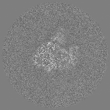
| 投影像・断面図 |
| ||||||||||||
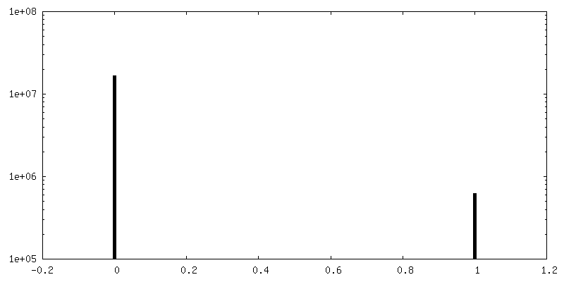
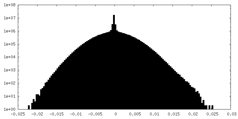
| 密度ヒストグラム |


-ハーフマップ: #2
| ファイル | emd_11999_half_map_2.map | ||||||||||||
|---|---|---|---|---|---|---|---|---|---|---|---|---|---|
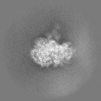
| 投影像・断面図 |
| ||||||||||||
| 密度ヒストグラム |


- 試料の構成要素
-全体 : 70S ribosome with chloramphenicol
| 全体 | 名称: 70S ribosome with chloramphenicol |
|---|---|
| 要素 |
|
-超分子 #1: 70S ribosome with chloramphenicol
| 超分子 | 名称: 70S ribosome with chloramphenicol / タイプ: complex / ID: 1 / 親要素: 0 |
|---|---|
| 由来(天然) | 生物種: Mycoplasma pneumoniae (バクテリア) |
| 分子量 | 実験値: 2.7 MDa |
-実験情報
-構造解析
| 手法 | クライオ電子顕微鏡法 |
|---|---|
 解析 解析 | サブトモグラム平均法 |
| 試料の集合状態 | particle |
-試料調製
| 緩衝液 | pH: 7 |
|---|---|
| 凍結 | 凍結剤: ETHANE |
- 電子顕微鏡法
| 顕微鏡 | FEI TITAN KRIOS |
|---|---|
| 電子線 | 加速電圧: 300 kV / 電子線源: FIELD EMISSION GUN |
| 電子光学系 | 照射モード: FLOOD BEAM / 撮影モード: BRIGHT FIELDBright-field microscopy |
| 撮影 | フィルム・検出器のモデル: GATAN K2 SUMMIT (4k x 4k) 平均電子線量: 120.0 e/Å2 |
| 実験機器 |  モデル: Titan Krios / 画像提供: FEI Company |
-画像解析
| 抽出 | トモグラム数: 65 / 使用した粒子像数: 24202 |
|---|---|
| 最終 角度割当 | タイプ: PROJECTION MATCHING |
| 最終 再構成 | 想定した対称性 - 点群: C1 (非対称) / アルゴリズム: FOURIER SPACE / 解像度のタイプ: BY AUTHOR / 解像度: 3.4 Å / 解像度の算出法: FSC 0.143 CUT-OFF / 使用したサブトモグラム数: 17890 |
| FSC曲線 (解像度の算出) | 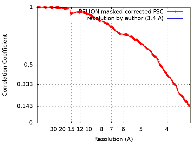 |
