 Movie
Movie Controller


+ Open data

- Basic information
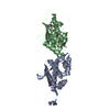
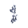
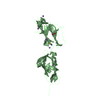
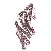
| Entry | Database: PDB / ID: 5.0E+37 | |||||||||
|---|---|---|---|---|---|---|---|---|---|---|
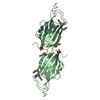
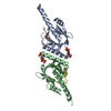
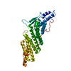
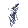
| Title | Redox protein from Chlamydomonas reinhardtii | |||||||||
 Components Components | EF-Hand domain-containing thioredoxin | |||||||||
 Keywords Keywords |  OXIDOREDUCTASE / Calredoxin / calcium- / redox- / Chlamydomonas reinhardtii OXIDOREDUCTASE / Calredoxin / calcium- / redox- / Chlamydomonas reinhardtii | |||||||||
| Function / homology |  Function and homology information Function and homology information | |||||||||
| Biological species |   Chlamydomonas reinhardtii (plant) Chlamydomonas reinhardtii (plant) | |||||||||
| Method | X-RAY DIFFRACTION / SYNCHROTRON / MOLECULAR REPLACEMENT / Resolution: 1.6 Å | |||||||||
 Authors Authors | Charoenwattansatien, R. / Hochmal, A.K. / Zinzius, K. / Muto, R. / Tanaka, H. / Hippler, M. / Kurisu, G. | |||||||||
| Funding support |  Japan, 2items Japan, 2items
| |||||||||
 Citation Citation | Journal: Nat Commun / Year: 2016 Title: Calredoxin represents a novel type of calcium-dependent sensor-responder connected to redox regulation in the chloroplast Authors: Hochmal, A.K. / Zinzius, K. / Charoenwattanasatien, R. / Gabelein, P. / Mutoh, R. / Tanaka, H. / Schulze, S. / Liu, G. / Scholz, M. / Nordhues, A. / Offenborn, J.N. / Petroutsos, D. / ...Authors: Hochmal, A.K. / Zinzius, K. / Charoenwattanasatien, R. / Gabelein, P. / Mutoh, R. / Tanaka, H. / Schulze, S. / Liu, G. / Scholz, M. / Nordhues, A. / Offenborn, J.N. / Petroutsos, D. / Finazzi, G. / Fufezan, C. / Huang, K. / Kurisu, G. / Hippler, M. | |||||||||
| History |
|
- Structure visualization
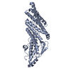
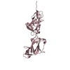
| Structure viewer | Molecule: MolmilJmol/JSmol |
|---|
- Downloads & links
-Download
| PDBx/mmCIF format | 5e37.cif.gz | 154.6 KB | Display | PDBx/mmCIF format |
|---|---|---|---|---|
| PDB format | pdb5e37.ent.gz | 124 KB | Display | PDB format |
| PDBx/mmJSON format | 5e37.json.gz | Tree view | PDBx/mmJSON format | |
| Others |  Other downloads Other downloads |
-Validation report
| Arichive directory | https://data.pdbj.org/pub/pdb/validation_reports/e3/5e37ftp://data.pdbj.org/pub/pdb/validation_reports/e3/5e37 | HTTPS FTP |
|---|
-Related structure data
| Similar structure data |
|---|
-Links
 PDBj
PDBj

- Assembly
| Deposited unit | 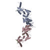
| ||||||||
|---|---|---|---|---|---|---|---|---|---|
| 1 | 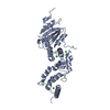
| ||||||||
| 2 | 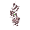
| ||||||||
| Unit cell |
|
-Components
| #1: Protein | Mass: 39950.398 Da / Num. of mol.: 2 Source method: isolated from a genetically manipulated source Source: (gene. exp.) Chlamydomonas reinhardtii (plant) / Gene: CHLREDRAFT_205510 / Production host:  Escherichia coli (E. coli) / Strain (production host): Rosetta / References: UniProt: A8IXH4*PLUS Escherichia coli (E. coli) / Strain (production host): Rosetta / References: UniProt: A8IXH4*PLUS#2: Chemical | ChemComp-CA /   Mass: 40.078 Da / Num. of mol.: 8 / Source method: obtained synthetically / Formula: Ca Mass: 40.078 Da / Num. of mol.: 8 / Source method: obtained synthetically / Formula: Ca#3: Water | ChemComp-HOH / | Water Mass: 18.015 Da / Num. of mol.: 849 / Source method: isolated from a natural source / Formula: H2O Mass: 18.015 Da / Num. of mol.: 849 / Source method: isolated from a natural source / Formula: H2OSequence details | THE SEQUENCE OF THIS PROTEIN WAS NOT AVAILABLE AT THE UNIPROT KNOWLEDGEBASE DATABASE (UNIPROTKB) AT ...THE SEQUENCE OF THIS PROTEIN WAS NOT AVAILABLE AT THE UNIPROT KNOWLEDGEB | |
|---|
-Experimental details
-Experiment
| Experiment | Method: X-RAY DIFFRACTION |
|---|
- Sample preparation
| Crystal | Density Matthews: 2.61 Å3/Da / Density % sol: 52.97 % |
|---|---|
| Crystal grow | Temperature: 293 K / Method: vapor diffusion, hanging drop / pH: 6 / Details: 1.5 M LiSO4 in 0.1 M MES-NaOH buffer pH 6.0 |
-Data collection
| Diffraction | Mean temperature: 100 K |
|---|---|
| Diffraction source | Source: SYNCHROTRON / Site: SPring-8 / Beamline: BL44XU / Wavelength: 0.9 Å |
| Detector | Type: RAYONIX MX300HE / Detector: CCD / Date: Apr 22, 2015 |
| Radiation | Protocol: SINGLE WAVELENGTH / Monochromatic (M) / Laue (L): M / Scattering type: x-ray |
| Radiation wavelength | Wavelength: 0.9 Å / Relative weight: 1 |
| Reflection | Resolution: 1.6→50 Å / Num. obs: 108097 / % possible obs: 99.7 % / Redundancy: 3.7 % / Rmerge(I) obs: 0.06 / Net I/σ(I): 35.3 |
| Reflection shell | Resolution: 1.6→1.63 Å / Redundancy: 3.6 % / Rmerge(I) obs: 0.558 / % possible all: 100 |
- Processing
| Software |
| ||||||||||||||||||||||||||||||||||||||||||||||||||||||||||||||||||||||||||||||||||||||||||||||||||||||||||||||||||||||||||||||||||||||||||||||||||||||||||||||||||||||||||||||||||||||
|---|---|---|---|---|---|---|---|---|---|---|---|---|---|---|---|---|---|---|---|---|---|---|---|---|---|---|---|---|---|---|---|---|---|---|---|---|---|---|---|---|---|---|---|---|---|---|---|---|---|---|---|---|---|---|---|---|---|---|---|---|---|---|---|---|---|---|---|---|---|---|---|---|---|---|---|---|---|---|---|---|---|---|---|---|---|---|---|---|---|---|---|---|---|---|---|---|---|---|---|---|---|---|---|---|---|---|---|---|---|---|---|---|---|---|---|---|---|---|---|---|---|---|---|---|---|---|---|---|---|---|---|---|---|---|---|---|---|---|---|---|---|---|---|---|---|---|---|---|---|---|---|---|---|---|---|---|---|---|---|---|---|---|---|---|---|---|---|---|---|---|---|---|---|---|---|---|---|---|---|---|---|---|---|
| Refinement | Method to determine structure: MOLECULAR REPLACEMENT / Resolution: 1.6→50 Å / Cor.coef. Fo:Fc: 0.955 / Cor.coef. Fo:Fc free: 0.943 / SU B: 1.794 / SU ML: 0.063 / Cross valid method: THROUGHOUT / ESU R: 0.086 / ESU R Free: 0.089 / Stereochemistry target values: MAXIMUM LIKELIHOOD / Details: HYDROGENS HAVE BEEN ADDED IN THE RIDING POSITIONS
| ||||||||||||||||||||||||||||||||||||||||||||||||||||||||||||||||||||||||||||||||||||||||||||||||||||||||||||||||||||||||||||||||||||||||||||||||||||||||||||||||||||||||||||||||||||||
| Solvent computation | Ion probe radii: 0.8 Å / Shrinkage radii: 0.8 Å / VDW probe radii: 1.2 Å / Solvent model: MASK | ||||||||||||||||||||||||||||||||||||||||||||||||||||||||||||||||||||||||||||||||||||||||||||||||||||||||||||||||||||||||||||||||||||||||||||||||||||||||||||||||||||||||||||||||||||||
| Displacement parameters | Biso mean: 25.518 Å2
| ||||||||||||||||||||||||||||||||||||||||||||||||||||||||||||||||||||||||||||||||||||||||||||||||||||||||||||||||||||||||||||||||||||||||||||||||||||||||||||||||||||||||||||||||||||||
| Refinement step | Cycle: 1 / Resolution: 1.6→50 Å
| ||||||||||||||||||||||||||||||||||||||||||||||||||||||||||||||||||||||||||||||||||||||||||||||||||||||||||||||||||||||||||||||||||||||||||||||||||||||||||||||||||||||||||||||||||||||
| Refine LS restraints |
|