 Movie
Movie Controller

[English] 日本語
 Yorodumi
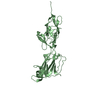
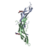
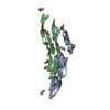
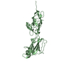
- PDB-5hbb: Crystal structure of shaft pilin spaA from Lactobacillus rhamnosu... -
+ Open data

- Basic information
| Entry | Database: PDB / ID: 5hbb | ||||||
|---|---|---|---|---|---|---|---|
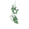
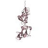
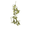
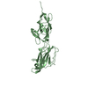
| Title | Crystal structure of shaft pilin spaA from Lactobacillus rhamnosus GG - E139A mutant | ||||||
 Components Components | Cell surface protein SpaA | ||||||
 Keywords Keywords | CELL ADHESION / Pilin / spaA / probiotic / isopeptide / SpaCBA pili / adhesin | ||||||
| Function / homology |  Function and homology information Function and homology informationGram-positive pilin subunit D1, N-terminal / Gram-positive pilin subunit D1, N-terminal domain / Prealbumin-like fold domain / Prealbumin-like fold domain / LPXTG cell wall anchor motif / Gram-positive cocci surface proteins LPxTG motif profile. / LPXTG cell wall anchor domain / Immunoglobulin-like fold / Immunoglobulins / Immunoglobulin-like ...Gram-positive pilin subunit D1, N-terminal / Gram-positive pilin subunit D1, N-terminal domain / Prealbumin-like fold domain / Prealbumin-like fold domain / LPXTG cell wall anchor motif / Gram-positive cocci surface proteins LPxTG motif profile. / LPXTG cell wall anchor domain / Immunoglobulin-like fold / Immunoglobulins / Immunoglobulin-like / Sandwich / Mainly Beta Similarity search - Domain/homology | ||||||
| Biological species |  Lactobacillus rhamnosus GG (bacteria) Lactobacillus rhamnosus GG (bacteria) | ||||||
| Method |  X-RAY DIFFRACTION / SYNCHROTRON / MOLECULAR REPLACEMENT / Resolution: 2.47 Å X-RAY DIFFRACTION / SYNCHROTRON / MOLECULAR REPLACEMENT / Resolution: 2.47 Å | ||||||
 Authors Authors | Chaurasia, P. / Pratap, S. / von Ossowski, I. / Palva, A. / Krishnan, V. | ||||||
| Funding support |  India, 1items India, 1items
| ||||||
 Citation Citation | Journal: Sci Rep / Year: 2016 Title: New insights about pilus formation in gut-adapted Lactobacillus rhamnosus GG from the crystal structure of the SpaA backbone-pilin subunit Authors: Chaurasia, P. / Pratap, S. / von Ossowski, I. / Palva, A. / Krishnan, V. | ||||||
| History |
|
- Structure visualization
| Structure viewer | Molecule: MolmilJmol/JSmol |
|---|
- Downloads & links
-Download
| PDBx/mmCIF format | 5hbb.cif.gz | 319.4 KB | Display | PDBx/mmCIF format |
|---|---|---|---|---|
| PDB format | pdb5hbb.ent.gz | 259.2 KB | Display | PDB format |
| PDBx/mmJSON format | 5hbb.json.gz | Tree view | PDBx/mmJSON format | |
| Others |  Other downloads Other downloads |
-Validation report
| Arichive directory | https://data.pdbj.org/pub/pdb/validation_reports/hb/5hbbftp://data.pdbj.org/pub/pdb/validation_reports/hb/5hbb | HTTPS FTP |
|---|
-Related structure data
| Related structure data |  5f44SC  5faaC 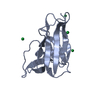 5fgrC  5fgsC 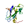 5fieC 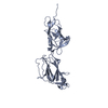 5hdlC  5htsC 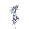 5j4mC S: Starting model for refinement C: citing same article ( |
|---|---|
| Similar structure data |


-Links
 PDBj
PDBj
- Assembly
| Deposited unit | 
| |||||||||||||||||||||||||||||||||||||||||||||||||||||||||||||||||||
|---|---|---|---|---|---|---|---|---|---|---|---|---|---|---|---|---|---|---|---|---|---|---|---|---|---|---|---|---|---|---|---|---|---|---|---|---|---|---|---|---|---|---|---|---|---|---|---|---|---|---|---|---|---|---|---|---|---|---|---|---|---|---|---|---|---|---|---|---|
| 1 | 
| |||||||||||||||||||||||||||||||||||||||||||||||||||||||||||||||||||
| 2 | 
| |||||||||||||||||||||||||||||||||||||||||||||||||||||||||||||||||||
| 3 | 
| |||||||||||||||||||||||||||||||||||||||||||||||||||||||||||||||||||
| Unit cell |
| |||||||||||||||||||||||||||||||||||||||||||||||||||||||||||||||||||
| Noncrystallographic symmetry (NCS) | NCS domain:
NCS domain segments: Component-ID: _ / Beg auth comp-ID: THR / Beg label comp-ID: THR / End auth comp-ID: LEU / End label comp-ID: LEU / Refine code: _
NCS ensembles :
|
-Components
| #1: Protein | Mass: 30665.463 Da / Num. of mol.: 3 / Fragment: UNP residues 35-302 / Mutation: E139A Source method: isolated from a genetically manipulated source Source: (gene. exp.) Lactobacillus rhamnosus GG (bacteria) / Strain: GG / Gene: LRHM_0426 / Production host: #2: Chemical |   Mass: 58.082 Da / Num. of mol.: 3 / Source method: obtained synthetically / Formula: CNS Mass: 58.082 Da / Num. of mol.: 3 / Source method: obtained synthetically / Formula: CNS#3: Chemical | ChemComp-NA /   Mass: 22.990 Da / Num. of mol.: 17 / Source method: obtained synthetically / Formula: Na Mass: 22.990 Da / Num. of mol.: 17 / Source method: obtained synthetically / Formula: Na#4: Chemical |   Mass: 62.068 Da / Num. of mol.: 3 / Source method: obtained synthetically / Formula: C2H6O2 Mass: 62.068 Da / Num. of mol.: 3 / Source method: obtained synthetically / Formula: C2H6O2#5: Water | ChemComp-HOH / |  Mass: 18.015 Da / Num. of mol.: 286 / Source method: isolated from a natural source / Formula: H2O Mass: 18.015 Da / Num. of mol.: 286 / Source method: isolated from a natural source / Formula: H2OHas protein modification | Y | |
|---|
-Experimental details
-Experiment
| Experiment | Method: X-RAY DIFFRACTION |
|---|
- Sample preparation
| Crystal | Density Matthews: 4.03 Å3/Da / Density % sol: 69.49 % |
|---|---|
| Crystal grow | Temperature: 277 K / Method: vapor diffusion, hanging drop / Details: 0.1M sodium thiocynate, 15% PEG3350 |
-Data collection
| Diffraction | Mean temperature: 100 K |
|---|---|
| Diffraction source | Source: SYNCHROTRON / Site: ESRF  / Beamline: BM14 / Wavelength: 0.95372 Å / Beamline: BM14 / Wavelength: 0.95372 Å |
| Detector | Type: MARMOSAIC 225 mm CCD / Detector: CCD / Date: Sep 24, 2015 |
| Radiation | Monochromator: Si / Protocol: SINGLE WAVELENGTH / Monochromatic (M) / Laue (L): M / Scattering type: x-ray |
| Radiation wavelength | Wavelength: 0.95372 Å / Relative weight: 1 |
| Reflection | Resolution: 2.47→111.8 Å / Num. obs: 44441 / % possible obs: 83.53 % / Redundancy: 5 % / Rmerge(I) obs: 0.08946 / Rsym value: 0.09943 / Net I/σ(I): 17.36 |
| Reflection shell | Resolution: 2.466→2.554 Å / Redundancy: 4 % / Rmerge(I) obs: 0.347 / Mean I/σ(I) obs: 3.84 / % possible all: 27.02 |
- Processing
| Software |
| ||||||||||||||||||||||||||||||||||||||||||||||||||||||||||||||||||||||||||||||||||||||||||||||||||||||||||||||||||||||||||||||||||||||||||||||||||||||||||||||||||||||||||||||||||||||
|---|---|---|---|---|---|---|---|---|---|---|---|---|---|---|---|---|---|---|---|---|---|---|---|---|---|---|---|---|---|---|---|---|---|---|---|---|---|---|---|---|---|---|---|---|---|---|---|---|---|---|---|---|---|---|---|---|---|---|---|---|---|---|---|---|---|---|---|---|---|---|---|---|---|---|---|---|---|---|---|---|---|---|---|---|---|---|---|---|---|---|---|---|---|---|---|---|---|---|---|---|---|---|---|---|---|---|---|---|---|---|---|---|---|---|---|---|---|---|---|---|---|---|---|---|---|---|---|---|---|---|---|---|---|---|---|---|---|---|---|---|---|---|---|---|---|---|---|---|---|---|---|---|---|---|---|---|---|---|---|---|---|---|---|---|---|---|---|---|---|---|---|---|---|---|---|---|---|---|---|---|---|---|---|
| Refinement | Method to determine structure: MOLECULAR REPLACEMENT Starting model: 5F44 Resolution: 2.47→111.8 Å / Cor.coef. Fo:Fc: 0.908 / Cor.coef. Fo:Fc free: 0.887 / SU B: 13.007 / SU ML: 0.154 / Cross valid method: THROUGHOUT / ESU R: 0.33 / ESU R Free: 0.224 / Stereochemistry target values: MAXIMUM LIKELIHOOD / Details: HYDROGENS HAVE BEEN ADDED IN THE RIDING POSITIONS
| ||||||||||||||||||||||||||||||||||||||||||||||||||||||||||||||||||||||||||||||||||||||||||||||||||||||||||||||||||||||||||||||||||||||||||||||||||||||||||||||||||||||||||||||||||||||
| Solvent computation | Ion probe radii: 0.8 Å / Shrinkage radii: 0.8 Å / VDW probe radii: 1 Å / Solvent model: MASK | ||||||||||||||||||||||||||||||||||||||||||||||||||||||||||||||||||||||||||||||||||||||||||||||||||||||||||||||||||||||||||||||||||||||||||||||||||||||||||||||||||||||||||||||||||||||
| Displacement parameters | Biso mean: 24.418 Å2
| ||||||||||||||||||||||||||||||||||||||||||||||||||||||||||||||||||||||||||||||||||||||||||||||||||||||||||||||||||||||||||||||||||||||||||||||||||||||||||||||||||||||||||||||||||||||
| Refinement step | Cycle: 1 / Resolution: 2.47→111.8 Å
| ||||||||||||||||||||||||||||||||||||||||||||||||||||||||||||||||||||||||||||||||||||||||||||||||||||||||||||||||||||||||||||||||||||||||||||||||||||||||||||||||||||||||||||||||||||||
| Refine LS restraints |
|