 Movie
Movie Controller

[English] 日本語
 Yorodumi
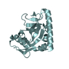
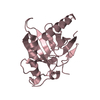
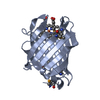
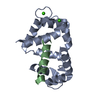
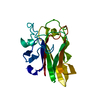
- PDB-5fie: Crystal structure of N-terminal domain of shaft pilin spaA from L... -
+ Open data

- Basic information
| Entry | Database: PDB / ID: 5fie | ||||||
|---|---|---|---|---|---|---|---|
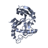
| Title | Crystal structure of N-terminal domain of shaft pilin spaA from Lactobacillus rhamnosus GG | ||||||
 Components Components | Cell surface protein SpaA | ||||||
 Keywords Keywords | CELL ADHESION / Pilin / spaA / probiotic / isopeptide / SpaCBA pili / adhesin | ||||||
| Function / homology |  Function and homology information Function and homology informationGram-positive pilin subunit D1, N-terminal / Gram-positive pilin subunit D1, N-terminal domain / Prealbumin-like fold domain / Prealbumin-like fold domain / LPXTG cell wall anchor motif / Gram-positive cocci surface proteins LPxTG motif profile. / LPXTG cell wall anchor domain / Immunoglobulin-like fold / Immunoglobulins / Immunoglobulin-like ...Gram-positive pilin subunit D1, N-terminal / Gram-positive pilin subunit D1, N-terminal domain / Prealbumin-like fold domain / Prealbumin-like fold domain / LPXTG cell wall anchor motif / Gram-positive cocci surface proteins LPxTG motif profile. / LPXTG cell wall anchor domain / Immunoglobulin-like fold / Immunoglobulins / Immunoglobulin-like / Sandwich / Mainly Beta Similarity search - Domain/homology | ||||||
| Biological species |  Lactobacillus rhamnosus GG (bacteria) Lactobacillus rhamnosus GG (bacteria) | ||||||
| Method |  X-RAY DIFFRACTION / MOLECULAR REPLACEMENT / Resolution: 2 Å X-RAY DIFFRACTION / MOLECULAR REPLACEMENT / Resolution: 2 Å | ||||||
 Authors Authors | Chaurasia, P. / Pratap, S. / von Ossowski, I. / Palva, A. / Krishnan, V. | ||||||
| Funding support |  India, 1items India, 1items
| ||||||
 Citation Citation | Journal: Sci Rep / Year: 2016 Title: New insights about pilus formation in gut-adapted Lactobacillus rhamnosus GG from the crystal structure of the SpaA backbone-pilin subunit Authors: Chaurasia, P. / Pratap, S. / von Ossowski, I. / Palva, A. / Krishnan, V. | ||||||
| History |
|
- Structure visualization
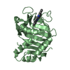
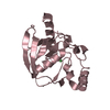
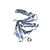
| Structure viewer | Molecule: MolmilJmol/JSmol |
|---|
- Downloads & links
-Download
| PDBx/mmCIF format | 5fie.cif.gz | 46.7 KB | Display | PDBx/mmCIF format |
|---|---|---|---|---|
| PDB format | pdb5fie.ent.gz | 30.2 KB | Display | PDB format |
| PDBx/mmJSON format | 5fie.json.gz | Tree view | PDBx/mmJSON format | |
| Others |  Other downloads Other downloads |
-Validation report
| Arichive directory | https://data.pdbj.org/pub/pdb/validation_reports/fi/5fieftp://data.pdbj.org/pub/pdb/validation_reports/fi/5fie | HTTPS FTP |
|---|
-Related structure data
| Related structure data |  5f44SC  5faaC 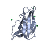 5fgrC  5fgsC  5hbbC 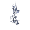 5hdlC  5htsC 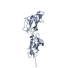 5j4mC S: Starting model for refinement C: citing same article ( |
|---|---|
| Similar structure data |

-Links
 PDBj
PDBj- Assembly
| Deposited unit | 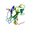
| ||||||||
|---|---|---|---|---|---|---|---|---|---|
| 1 |
| ||||||||
| Unit cell |
| ||||||||
| Components on special symmetry positions |
|
-Components
| #1: Protein | Mass: 16716.312 Da / Num. of mol.: 1 / Fragment: UNP residues 35-180 Source method: isolated from a genetically manipulated source Source: (gene. exp.) Lactobacillus rhamnosus GG (bacteria) / Strain: GG / Gene: LRHM_0426 / Production host: |
|---|---|
| #2: Chemical | ChemComp-NA /   Mass: 22.990 Da / Num. of mol.: 1 / Source method: obtained synthetically / Formula: Na Mass: 22.990 Da / Num. of mol.: 1 / Source method: obtained synthetically / Formula: Na |
| #3: Water | ChemComp-HOH /  Mass: 18.015 Da / Num. of mol.: 123 / Source method: isolated from a natural source / Formula: H2O Mass: 18.015 Da / Num. of mol.: 123 / Source method: isolated from a natural source / Formula: H2O |
-Experimental details
-Experiment
| Experiment | Method: X-RAY DIFFRACTION |
|---|
- Sample preparation
| Crystal | Density Matthews: 3.09 Å3/Da / Density % sol: 60.21 % |
|---|---|
| Crystal grow | Temperature: 295 K / Method: vapor diffusion, hanging drop / Details: 0.2M sodium formate, 20% PEG 3350 |
-Data collection
| Diffraction | Mean temperature: 100 K |
|---|---|
| Diffraction source | Source: ROTATING ANODE / Type: RIGAKU FR-E+ SUPERBRIGHT / Wavelength: 1.54178 Å |
| Detector | Type: RIGAKU RAXIS IV++ / Detector: IMAGE PLATE / Date: Sep 10, 2015 |
| Radiation | Protocol: SINGLE WAVELENGTH / Monochromatic (M) / Laue (L): M / Scattering type: x-ray |
| Radiation wavelength | Wavelength: 1.54178 Å / Relative weight: 1 |
| Reflection | Resolution: 2→48.89 Å / Num. obs: 13657 / % possible obs: 99.9 % / Redundancy: 6.9 % / Rmerge(I) obs: 0.135 / Net I/σ(I): 11.7 |
| Reflection shell | Resolution: 2→2.19 Å / Redundancy: 3.6 % / Rmerge(I) obs: 0.645 / Mean I/σ(I) obs: 3.7 / % possible all: 99.8 |
- Processing
| Software |
| ||||||||||||||||||||||||||||||||||||||||||
|---|---|---|---|---|---|---|---|---|---|---|---|---|---|---|---|---|---|---|---|---|---|---|---|---|---|---|---|---|---|---|---|---|---|---|---|---|---|---|---|---|---|---|---|
| Refinement | Method to determine structure: MOLECULAR REPLACEMENT Starting model: 5F44 Resolution: 2→44.307 Å / SU ML: 0.18 / Cross valid method: FREE R-VALUE / σ(F): 1.34 / Phase error: 23.71 / Stereochemistry target values: ML
| ||||||||||||||||||||||||||||||||||||||||||
| Solvent computation | Shrinkage radii: 0.9 Å / VDW probe radii: 1.11 Å / Solvent model: FLAT BULK SOLVENT MODEL | ||||||||||||||||||||||||||||||||||||||||||
| Refinement step | Cycle: LAST / Resolution: 2→44.307 Å
| ||||||||||||||||||||||||||||||||||||||||||
| Refine LS restraints |
| ||||||||||||||||||||||||||||||||||||||||||
| LS refinement shell |
|
