Resolution: 2.55→30 Å / Cor.coef. Fo:Fc: 0.94 / Cor.coef. Fo:Fc free: 0.916 / SU B: 19.14 / SU ML: 0.217 / Cross valid method: THROUGHOUT / ESU R: 0.533 / ESU R Free: 0.32 / Stereochemistry target values: MAXIMUM LIKELIHOOD / Details: HYDROGENS HAVE BEEN ADDED IN THE RIDING POSITIONS
Rfactor
Num. reflection
% reflection
Selection details
Rfree
0.27386
783
5.1 %
RANDOM
Rwork
0.20827
-
-
-
obs
0.21167
14704
96.43 %
-
all
-
16079
-
-
Solvent computation
Ion probe radii: 0.8 Å / Shrinkage radii: 0.8 Å / VDW probe radii: 1.4 Å / Solvent model: MASK
Displacement parameters
Biso mean: 32.059 Å2
Baniso -1
Baniso -2
Baniso -3
1-
-1.28 Å2
0 Å2
1.05 Å2
2-
-
3.3 Å2
0 Å2
3-
-
-
-0.91 Å2
Refinement step
Cycle: LAST / Resolution: 2.55→30 Å
Protein
Nucleic acid
Ligand
Solvent
Total
Num. atoms
2828
0
0
82
2910
Refine LS restraints
Refine-ID
Type
Dev ideal
Dev ideal target
Number
X-RAY DIFFRACTION
r_bond_refined_d
0.016
0.022
2884
X-RAY DIFFRACTION
r_bond_other_d
X-RAY DIFFRACTION
r_angle_refined_deg
1.544
1.969
3896
X-RAY DIFFRACTION
r_angle_other_deg
X-RAY DIFFRACTION
r_dihedral_angle_1_deg
6.901
5
363
X-RAY DIFFRACTION
r_dihedral_angle_2_deg
36.844
25.267
131
X-RAY DIFFRACTION
r_dihedral_angle_3_deg
20.348
15
528
X-RAY DIFFRACTION
r_dihedral_angle_4_deg
22.113
15
11
X-RAY DIFFRACTION
r_chiral_restr
0.106
0.2
438
X-RAY DIFFRACTION
r_gen_planes_refined
0.005
0.02
2155
X-RAY DIFFRACTION
r_gen_planes_other
X-RAY DIFFRACTION
r_nbd_refined
0.228
0.2
1361
X-RAY DIFFRACTION
r_nbd_other
X-RAY DIFFRACTION
r_nbtor_refined
0.312
0.2
1961
X-RAY DIFFRACTION
r_nbtor_other
X-RAY DIFFRACTION
r_xyhbond_nbd_refined
0.16
0.2
102
X-RAY DIFFRACTION
r_xyhbond_nbd_other
X-RAY DIFFRACTION
r_metal_ion_refined
X-RAY DIFFRACTION
r_metal_ion_other
X-RAY DIFFRACTION
r_symmetry_vdw_refined
0.141
0.2
17
X-RAY DIFFRACTION
r_symmetry_vdw_other
X-RAY DIFFRACTION
r_symmetry_hbond_refined
0.198
0.2
5
X-RAY DIFFRACTION
r_symmetry_hbond_other
X-RAY DIFFRACTION
r_symmetry_metal_ion_refined
X-RAY DIFFRACTION
r_symmetry_metal_ion_other
X-RAY DIFFRACTION
r_mcbond_it
0.755
1.5
1846
X-RAY DIFFRACTION
r_mcbond_other
X-RAY DIFFRACTION
r_mcangle_it
1.253
2
2887
X-RAY DIFFRACTION
r_scbond_it
1.78
3
1159
X-RAY DIFFRACTION
r_scangle_it
2.82
4.5
1006
X-RAY DIFFRACTION
r_rigid_bond_restr
X-RAY DIFFRACTION
r_sphericity_free
X-RAY DIFFRACTION
r_sphericity_bonded
LS refinement shell
Resolution: 2.549→2.615 Å / Total num. of bins used: 20
Rfactor
Num. reflection
% reflection
Rfree
0.323
48
-
Rwork
0.289
859
-
obs
-
-
77.46 %
Refinement TLS params.
Method: refined / Refine-ID: X-RAY DIFFRACTION
ID
L11 (°2)
L12 (°2)
L13 (°2)
L22 (°2)
L23 (°2)
L33 (°2)
S11 (Å °)
S12 (Å °)
S13 (Å °)
S21 (Å °)
S22 (Å °)
S23 (Å °)
S31 (Å °)
S32 (Å °)
S33 (Å °)
T11 (Å2)
T12 (Å2)
T13 (Å2)
T22 (Å2)
T23 (Å2)
T33 (Å2)
Origin x (Å)
Origin y (Å)
Origin z (Å)
1
19.6737
6.7744
-4.3723
2.5736
-1.7487
3.6439
-0.4958
0.0794
-0.8158
0.0107
0.0269
-0.0986
0.5955
-0.2716
0.4689
0.265
-0.1018
0.0256
0.1851
-0.0671
0.241
14.6001
20.3462
24.5862
2
27.1824
4.048
-2.7682
0.6132
-0.517
1.3431
1.0747
0.3194
0.8322
0.5251
-0.7371
1.8113
-2.2744
-1.2451
-0.3376
0.5369
-0.0038
-0.0004
0.5362
0.0015
0.5372
-15.9165
28.4233
22.1912
3
15.9299
2.9643
-2.9387
5.0716
-2.7557
4.4404
-0.6135
0.9044
-0.4868
-0.5573
0.36
-0.0094
0.9035
-0.3771
0.2535
0.1315
-0.1489
-0.0097
0.243
-0.1607
0.1267
5.1737
22.0315
17.7907
4
7.5555
8.359
-1.5774
19.4043
-12.1111
10.9093
0.3888
-1.0288
1.9688
3.4554
-0.0763
1.3447
-1.5783
-1.5994
-0.3125
0.4853
-0.0013
-0.0061
0.4851
0.0014
0.4884
-5.5243
35.7201
28.6481
5
4.3429
2.0082
-6.1562
7.6627
-1.4139
9.0316
0.3718
-1.8647
-0.9481
0.7099
-0.6731
-0.1941
0.4926
0.2683
0.3013
0.2126
-0.2208
0.006
0.4412
0.0313
0.1758
11.0618
21.0305
36.1779
6
6.8241
2.6474
-6.4688
1.027
-2.5096
6.132
0.6165
-1.0914
-1.0838
-0.4036
-1.1042
0.4006
1.4007
0.4923
0.4876
0.4008
-0.0522
0.0323
0.4005
-0.0308
0.395
7.1306
24.6946
37.3303
7
6.4598
0.0459
-3.1552
2.4346
0.106
3.7577
-0.3217
0.8026
-0.104
-0.3065
0.0146
0.0379
0.2423
-0.4192
0.3071
0.1897
-0.1091
-0.0576
0.2662
-0.0854
0.1617
18.4217
25.3011
15.1381
8
4.7553
0.1426
-1.052
3.6331
-0.7601
4.8106
0.0658
0.1759
0.6554
0.1053
0.0189
0.1237
-0.5236
-0.2586
-0.0847
0.1154
-0.0395
-0.0407
0.1818
-0.0652
0.2371
17.973
34.4269
21.4649
9
5.5339
1.2466
-1.9607
1.7111
-0.3847
3.9475
-0.0225
0.0919
0.2889
0.0225
-0.0383
0.2108
-0.0865
-0.1086
0.0608
0.2394
-0.0226
-0.0498
0.1991
-0.0138
0.2316
27.7865
33.4903
20.2404
10
2.696
-0.4689
-2.7313
0.6036
1.9568
6.973
-0.114
-0.5279
-0.2488
0.2699
-0.2451
-0.0095
0.3917
-0.3765
0.3591
0.1732
-0.0446
-0.0046
0.2189
0.0422
0.1641
31.6007
26.067
31.3345
11
2.8851
0.5029
-1.7214
1.7892
-1.0628
1.5696
-0.0389
0.0298
-0.0001
-0.0535
-0.0092
0.0082
0.0848
0.0328
0.0481
0.1872
-0.0236
-0.0235
0.2339
-0.0194
0.1182
49.1872
32.222
13.8129
12
6.2733
0.558
-2.1341
0.7677
-0.3947
1.3979
0.0143
0.4159
0.1126
-0.0728
-0.0152
0.1339
0.098
-0.1181
0.0008
0.1825
-0.0508
-0.0577
0.1417
-0.0447
0.0678
37.6024
31.772
13.4729
Refinement TLS group
Refine-ID: X-RAY DIFFRACTION / Selection: ALL / Auth asym-ID: A / Label asym-ID: A
ID
Refine TLS-ID
Auth seq-ID
Label seq-ID
1
1
1 - 17
22 - 38
2
2
18 - 45
39 - 66
3
3
46 - 74
67 - 95
4
4
75 - 89
96 - 110
5
5
90 - 105
111 - 126
6
6
106 - 116
127 - 137
7
7
117 - 152
138 - 173
8
8
153 - 165
174 - 186
9
9
166 - 230
187 - 251
10
10
231 - 247
252 - 268
11
11
248 - 290
269 - 311
12
12
291 - 358
312 - 379
+
About Yorodumi
-
News
-
Feb 9, 2022. New format data for meta-information of EMDB entries
New format data for meta-information of EMDB entries
Version 3 of the EMDB header file is now the official format.
The previous official version 1.9 will be removed from the archive.
In the structure databanks used in Yorodumi, some data are registered as the other names, "COVID-19 virus" and "2019-nCoV". Here are the details of the virus and the list of structure data.
Jan 31, 2019. EMDB accession codes are about to change! (news from PDBe EMDB page)
EMDB accession codes are about to change! (news from PDBe EMDB page)
The allocation of 4 digits for EMDB accession codes will soon come to an end. Whilst these codes will remain in use, new EMDB accession codes will include an additional digit and will expand incrementally as the available range of codes is exhausted. The current 4-digit format prefixed with “EMD-” (i.e. EMD-XXXX) will advance to a 5-digit format (i.e. EMD-XXXXX), and so on. It is currently estimated that the 4-digit codes will be depleted around Spring 2019, at which point the 5-digit format will come into force.
The EM Navigator/Yorodumi systems omit the EMD- prefix.
Related info.:Q: What is EMD? / ID/Accession-code notation in Yorodumi/EM Navigator
Yorodumi is a browser for structure data from EMDB, PDB, SASBDB, etc.
This page is also the successor to EM Navigator detail page, and also detail information page/front-end page for Omokage search.
The word "yorodu" (or yorozu) is an old Japanese word meaning "ten thousand". "mi" (miru) is to see.
Related info.:EMDB / PDB / SASBDB / Comparison of 3 databanks / Yorodumi Search / Aug 31, 2016. New EM Navigator & Yorodumi / Yorodumi Papers / Jmol/JSmol / Function and homology information / Changes in new EM Navigator and Yorodumi
 Movie
Movie Controller


 Open data
 Basic information
 Components
 Keywords
 Function and homology information
 Homo sapiens (human)
 X-RAY DIFFRACTION /
  Authors
 Citation
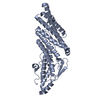
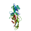
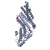
 Structure visualization
 Downloads & links
 Other downloads


 PDBj
PDBj


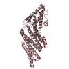
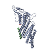
 Assembly
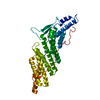

 Mass: 18.015 Da / Num. of mol.: 79 / Source method: isolated from a natural source / Formula: H2O
 Sample preparation
 Processing