 Movie
Movie Controller

[English] 日本語
 Yorodumi
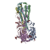
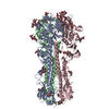
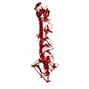
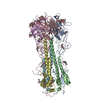
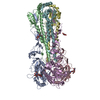
- PDB-4i78: Crystal structure of a subtype H17 hemagglutinin homologue from A... -
+ Open data

- Basic information
| Entry | Database: PDB / ID: 4i78 | |||||||||
|---|---|---|---|---|---|---|---|---|---|---|
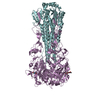
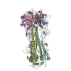
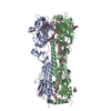
| Title | Crystal structure of a subtype H17 hemagglutinin homologue from A/little yellow-shouldered bat/Guatemala/060/2010 (H17N10) | |||||||||
 Components Components |
| |||||||||
 Keywords Keywords |  VIRAL PROTEIN / influenza vrus / hemagglutinin homologue / H17 / ectodomain VIRAL PROTEIN / influenza vrus / hemagglutinin homologue / H17 / ectodomain | |||||||||
| Function / homology |  Function and homology informationviral budding from plasma membrane / clathrin-dependent endocytosis of virus by host cell / host cell surface receptor binding / apical plasma membrane / fusion of virus membrane with host plasma membrane / fusion of virus membrane with host endosome membrane / viral envelope / virion attachment to host cell / host cell plasma membrane / virion membrane Function and homology informationviral budding from plasma membrane / clathrin-dependent endocytosis of virus by host cell / host cell surface receptor binding / apical plasma membrane / fusion of virus membrane with host plasma membrane / fusion of virus membrane with host endosome membrane / viral envelope / virion attachment to host cell / host cell plasma membrane / virion membraneSimilarity search - Function | |||||||||
| Biological species |   Influenza A virus Influenza A virus | |||||||||
| Method | X-RAY DIFFRACTION / SYNCHROTRON / MOLECULAR REPLACEMENT / Resolution: 3.18 Å | |||||||||
 Authors Authors | Zhu, X. / Wilson, I.A. | |||||||||
 Citation Citation | Journal: Proc.Natl.Acad.Sci.USA / Year: 2013 Title: Hemagglutinin homologue from H17N10 bat influenza virus exhibits divergent receptor-binding and pH-dependent fusion activities. Authors: Zhu, X. / Yu, W. / McBride, R. / Li, Y. / Chen, L.M. / Donis, R.O. / Tong, S. / Paulson, J.C. / Wilson, I.A. | |||||||||
| History |
|
- Structure visualization
| Structure viewer | Molecule: MolmilJmol/JSmol |
|---|
- Downloads & links
-Download
| PDBx/mmCIF format | 4i78.cif.gz | 204.4 KB | Display | PDBx/mmCIF format |
|---|---|---|---|---|
| PDB format | pdb4i78.ent.gz | 164 KB | Display | PDB format |
| PDBx/mmJSON format | 4i78.json.gz | Tree view | PDBx/mmJSON format | |
| Others |  Other downloads Other downloads |
-Validation report
| Arichive directory | https://data.pdbj.org/pub/pdb/validation_reports/i7/4i78ftp://data.pdbj.org/pub/pdb/validation_reports/i7/4i78 | HTTPS FTP |
|---|
-Related structure data
| Related structure data | 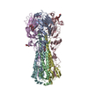 3ubqS S: Starting model for refinement |
|---|---|
| Similar structure data |
-Links
 PDBj
PDBj


- Assembly
| Deposited unit | 
| ||||||||
|---|---|---|---|---|---|---|---|---|---|
| 1 | 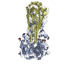
| ||||||||
| 2 | 
| ||||||||
| Unit cell |
|
-Components
| #1: Protein | Mass: 36696.008 Da / Num. of mol.: 2 Source method: isolated from a genetically manipulated source Source: (gene. exp.) Influenza A virusStrain: A/little yellow-shouldered bat/Guatemala/060/2010 (H17N10) Gene: HA / Plasmid: PFASTBACHT-A / Production host:  TRICHOPLUSIA NI (cabbage looper) / Strain (production host): HI5 / References: UniProt: H6QM93 TRICHOPLUSIA NI (cabbage looper) / Strain (production host): HI5 / References: UniProt: H6QM93#2: Protein | Mass: 20749.148 Da / Num. of mol.: 2 / Mutation: A47G Source method: isolated from a genetically manipulated source Source: (gene. exp.) Influenza A virusStrain: A/little yellow-shouldered bat/Guatemala/060/2010 (H17N10) Gene: HA / Plasmid: PFASTBACHT-A / Production host: TRICHOPLUSIA NI (cabbage looper) / Strain (production host): HI5 / References: UniProt: H6QM93#3: Polysaccharide | alpha-L-fucopyranose-(1-6)-2-acetamido-2-deoxy-beta-D-glucopyranose | / Mass: 367.349 Da / Num. of mol.: 1Source method: isolated from a genetically manipulated source #4: Sugar | ChemComp-NAG / | N-Acetylglucosamine  Type: D-saccharide, beta linking / Mass: 221.208 Da / Num. of mol.: 1 Type: D-saccharide, beta linking / Mass: 221.208 Da / Num. of mol.: 1Source method: isolated from a genetically manipulated source Formula: C8H15NO6 |
|---|
-Experimental details
-Experiment
| Experiment | Method: X-RAY DIFFRACTION |
|---|
- Sample preparation
| Crystal | Density Matthews: 3.24 Å3/Da / Density % sol: 62.01 % |
|---|---|
| Crystal grow | Temperature: 295 K / Method: vapor diffusion, sitting drop / pH: 5.5 Details: 0.1 M sodium citrate, pH 5.5, 1M LiCl2, and 15% PEG6000, VAPOR DIFFUSION, SITTING DROP, temperature 295K |
-Data collection
| Diffraction | Mean temperature: 100 K |
|---|---|
| Diffraction source | Source: SYNCHROTRON / Site: CLSI  / Beamline: 08ID-1 / Wavelength: 0.97949 Å / Beamline: 08ID-1 / Wavelength: 0.97949 Å |
| Detector | Type: RAYONIX MX-300 / Detector: CCD / Date: Sep 11, 2012 |
| Radiation | Monochromator: Vertical Focusing Mirror: ultra-low expansion (ULE) titanium siliicate flat mirror with Pt, Uncoated, and Pd strips Protocol: SINGLE WAVELENGTH / Monochromatic (M) / Laue (L): M / Scattering type: x-ray |
| Radiation wavelength | Wavelength: 0.97949 Å / Relative weight: 1 |
| Reflection | Resolution: 3.18→50 Å / Num. obs: 24997 / % possible obs: 97.6 % / Observed criterion σ(I): -3 |
| Reflection shell | Resolution: 3.18→3.27 Å / % possible all: 91.2 |
- Processing
| Software |
| ||||||||||||||||||||||||||||||||||||||||||||||||||||||||||||||||||||||||||||||||||||||||||||||||||||||||||||||||||||||||||||||||||||||||||||||||||||||||||||||||||||||||||
|---|---|---|---|---|---|---|---|---|---|---|---|---|---|---|---|---|---|---|---|---|---|---|---|---|---|---|---|---|---|---|---|---|---|---|---|---|---|---|---|---|---|---|---|---|---|---|---|---|---|---|---|---|---|---|---|---|---|---|---|---|---|---|---|---|---|---|---|---|---|---|---|---|---|---|---|---|---|---|---|---|---|---|---|---|---|---|---|---|---|---|---|---|---|---|---|---|---|---|---|---|---|---|---|---|---|---|---|---|---|---|---|---|---|---|---|---|---|---|---|---|---|---|---|---|---|---|---|---|---|---|---|---|---|---|---|---|---|---|---|---|---|---|---|---|---|---|---|---|---|---|---|---|---|---|---|---|---|---|---|---|---|---|---|---|---|---|---|---|---|---|---|
| Refinement | Method to determine structure: MOLECULAR REPLACEMENT Starting model: PDB ENTRY 3UBQ Resolution: 3.18→50 Å / Cor.coef. Fo:Fc: 0.924 / Cor.coef. Fo:Fc free: 0.878 / SU B: 20.254 / SU ML: 0.346 / Cross valid method: THROUGHOUT / ESU R Free: 0.485 / Stereochemistry target values: MAXIMUM LIKELIHOOD / Details: HYDROGENS HAVE BEEN USED IF PRESENT IN THE INPUT
| ||||||||||||||||||||||||||||||||||||||||||||||||||||||||||||||||||||||||||||||||||||||||||||||||||||||||||||||||||||||||||||||||||||||||||||||||||||||||||||||||||||||||||
| Solvent computation | Ion probe radii: 0.8 Å / Shrinkage radii: 0.8 Å / VDW probe radii: 1.2 Å / Solvent model: MASK | ||||||||||||||||||||||||||||||||||||||||||||||||||||||||||||||||||||||||||||||||||||||||||||||||||||||||||||||||||||||||||||||||||||||||||||||||||||||||||||||||||||||||||
| Displacement parameters | Biso mean: 64.649 Å2
| ||||||||||||||||||||||||||||||||||||||||||||||||||||||||||||||||||||||||||||||||||||||||||||||||||||||||||||||||||||||||||||||||||||||||||||||||||||||||||||||||||||||||||
| Refinement step | Cycle: LAST / Resolution: 3.18→50 Å
| ||||||||||||||||||||||||||||||||||||||||||||||||||||||||||||||||||||||||||||||||||||||||||||||||||||||||||||||||||||||||||||||||||||||||||||||||||||||||||||||||||||||||||
| Refine LS restraints |
| ||||||||||||||||||||||||||||||||||||||||||||||||||||||||||||||||||||||||||||||||||||||||||||||||||||||||||||||||||||||||||||||||||||||||||||||||||||||||||||||||||||||||||
| LS refinement shell | Resolution: 3.181→3.264 Å / Total num. of bins used: 20
|
