 Movie
Movie Controller

[English] 日本語
 Yorodumi
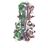
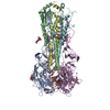
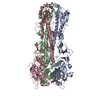
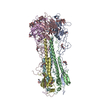
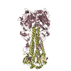
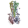
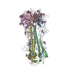
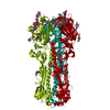
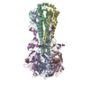
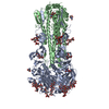
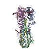
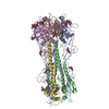
- PDB-6v46: The crystal structure of hemagglutinin from A/turkey/Ontario/6118... -
+ Open data

- Basic information
| Entry | Database: PDB / ID: 6v46 | |||||||||
|---|---|---|---|---|---|---|---|---|---|---|
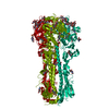
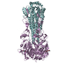
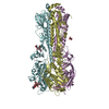
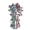
| Title | The crystal structure of hemagglutinin from A/turkey/Ontario/6118/1968 (H8N4) | |||||||||
 Components Components | (Hemagglutinin ...) x 2 | |||||||||
 Keywords Keywords | VIRAL PROTEIN / Influenza / avian / H8 | |||||||||
| Function / homology |  Function and homology information Function and homology informationviral budding from plasma membrane / clathrin-dependent endocytosis of virus by host cell / apical plasma membrane / host cell surface receptor binding / fusion of virus membrane with host plasma membrane / fusion of virus membrane with host endosome membrane / viral envelope / virion attachment to host cell / host cell plasma membrane / virion membrane / membrane Similarity search - Function | |||||||||
| Biological species |   Influenza A virus Influenza A virus | |||||||||
| Method |  X-RAY DIFFRACTION / SYNCHROTRON / MOLECULAR REPLACEMENT / Resolution: 2.248 Å X-RAY DIFFRACTION / SYNCHROTRON / MOLECULAR REPLACEMENT / Resolution: 2.248 Å | |||||||||
 Authors Authors | Yang, H. / Stevens, J. | |||||||||
 Citation Citation | Journal: Heliyon / Year: 2020 Title: Molecular characterization and three-dimensional structures of avian H8, H11, H14, H15 and swine H4 influenza virus hemagglutinins Authors: Yang, H. / Carney, P.J. / Chang, J. / Stevens, J. | |||||||||
| History |
|
- Structure visualization
| Structure viewer | Molecule: MolmilJmol/JSmol |
|---|
- Downloads & links
-Download
| PDBx/mmCIF format | 6v46.cif.gz | 601 KB | Display | PDBx/mmCIF format |
|---|---|---|---|---|
| PDB format | pdb6v46.ent.gz | 496.5 KB | Display | PDB format |
| PDBx/mmJSON format | 6v46.json.gz | Tree view | PDBx/mmJSON format | |
| Others |  Other downloads Other downloads |
-Validation report
| Arichive directory | https://data.pdbj.org/pub/pdb/validation_reports/v4/6v46ftp://data.pdbj.org/pub/pdb/validation_reports/v4/6v46 | HTTPS FTP |
|---|
-Related structure data
| Related structure data | 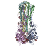 6v44C 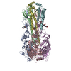 6v47C 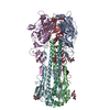 6v48C  6v49C 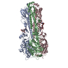 4wsrS S: Starting model for refinement C: citing same article ( |
|---|---|
| Similar structure data |

-Links
 PDBj
PDBj


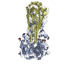
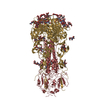
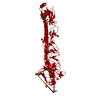
- Assembly
| Deposited unit | 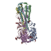
| |||||||||||||||||||||||||||||||||||||||||||||||||||||||||||||||||||||||||||||||||||||||
|---|---|---|---|---|---|---|---|---|---|---|---|---|---|---|---|---|---|---|---|---|---|---|---|---|---|---|---|---|---|---|---|---|---|---|---|---|---|---|---|---|---|---|---|---|---|---|---|---|---|---|---|---|---|---|---|---|---|---|---|---|---|---|---|---|---|---|---|---|---|---|---|---|---|---|---|---|---|---|---|---|---|---|---|---|---|---|---|---|
| 1 |
| |||||||||||||||||||||||||||||||||||||||||||||||||||||||||||||||||||||||||||||||||||||||
| Unit cell |
| |||||||||||||||||||||||||||||||||||||||||||||||||||||||||||||||||||||||||||||||||||||||
| Noncrystallographic symmetry (NCS) | NCS domain:
NCS domain segments: Component-ID: 1 / End auth comp-ID: SER / End label comp-ID: SER
NCS ensembles :
|
-Components
-Hemagglutinin ... , 2 types, 6 molecules ACEBDF
| #1: Protein | Mass: 37294.805 Da / Num. of mol.: 3 Source method: isolated from a genetically manipulated source Source: (gene. exp.) Influenza A virus (strain A/Turkey/Ontario/6118/1968 H8N4)Strain: A/Turkey/Ontario/6118/1968 H8N4 / Gene: HA / Production host:  Trichoplusia ni (cabbage looper) / References: UniProt: F2P175, UniProt: P03456*PLUS Trichoplusia ni (cabbage looper) / References: UniProt: F2P175, UniProt: P03456*PLUS#2: Protein | Mass: 20770.014 Da / Num. of mol.: 3 Source method: isolated from a genetically manipulated source Source: (gene. exp.) Influenza A virus (strain A/Turkey/Ontario/6118/1968 H8N4)Strain: A/Turkey/Ontario/6118/1968 H8N4 / Gene: HA / Production host: Trichoplusia ni (cabbage looper) / References: UniProt: F2P175, UniProt: P03456*PLUS |
|---|
-Sugars , 3 types, 9 molecules 
| #3: Polysaccharide | Source method: isolated from a genetically manipulated source #4: Polysaccharide | Source method: isolated from a genetically manipulated source #5: Sugar |  Type: D-saccharide, beta linking / Mass: 221.208 Da / Num. of mol.: 3 Type: D-saccharide, beta linking / Mass: 221.208 Da / Num. of mol.: 3Source method: isolated from a genetically manipulated source Formula: C8H15NO6 |
|---|
-Non-polymers , 1 types, 557 molecules 
| #6: Water | ChemComp-HOH / Mass: 18.015 Da / Num. of mol.: 557 / Source method: isolated from a natural source / Formula: H2O |
|---|
-Details
| Has ligand of interest | N |
|---|---|
| Has protein modification | Y |
-Experimental details
-Experiment
| Experiment | Method: X-RAY DIFFRACTION / Number of used crystals: 1 |
|---|
- Sample preparation
| Crystal | Density Matthews: 2.87 Å3/Da / Density % sol: 57.22 % |
|---|---|
| Crystal grow | Temperature: 293 K / Method: microbatch Details: 0.05M Magnesium Chloride, 0.1M HEPES:NaOH pH7.5, 30% (v/v) PEG 550 MME |
-Data collection
| Diffraction | Mean temperature: 100 K / Serial crystal experiment: N |
|---|---|
| Diffraction source | Source: SYNCHROTRON / Site: APS  / Beamline: 22-ID / Wavelength: 1 Å / Beamline: 22-ID / Wavelength: 1 Å |
| Detector | Type: MARMOSAIC 325 mm CCD / Detector: CCD / Date: Nov 14, 2011 |
| Radiation | Protocol: SINGLE WAVELENGTH / Monochromatic (M) / Laue (L): M / Scattering type: x-ray |
| Radiation wavelength | Wavelength: 1 Å / Relative weight: 1 |
| Reflection | Resolution: 2.248→50 Å / Num. obs: 91049 / % possible obs: 98.2 % / Redundancy: 3.8 % / Rsym value: 0.085 / Net I/σ(I): 3.8 |
| Reflection shell | Resolution: 2.25→2.33 Å / Num. unique obs: 4561 / Rsym value: 0.769 |
- Processing
| Software |
| ||||||||||||||||||||||||||||||||||||||||||||||||||||||||||||||||||||||||||||||||||||||||||||||||||||||||||||||||||||||||||||||||||||||||||||||||||||||||||||||||||||||||||||||||||||||||||
|---|---|---|---|---|---|---|---|---|---|---|---|---|---|---|---|---|---|---|---|---|---|---|---|---|---|---|---|---|---|---|---|---|---|---|---|---|---|---|---|---|---|---|---|---|---|---|---|---|---|---|---|---|---|---|---|---|---|---|---|---|---|---|---|---|---|---|---|---|---|---|---|---|---|---|---|---|---|---|---|---|---|---|---|---|---|---|---|---|---|---|---|---|---|---|---|---|---|---|---|---|---|---|---|---|---|---|---|---|---|---|---|---|---|---|---|---|---|---|---|---|---|---|---|---|---|---|---|---|---|---|---|---|---|---|---|---|---|---|---|---|---|---|---|---|---|---|---|---|---|---|---|---|---|---|---|---|---|---|---|---|---|---|---|---|---|---|---|---|---|---|---|---|---|---|---|---|---|---|---|---|---|---|---|---|---|---|---|
| Refinement | Method to determine structure: MOLECULAR REPLACEMENT Starting model: 4WSR Resolution: 2.248→45.412 Å / SU ML: 0.26 / Cross valid method: THROUGHOUT / σ(F): 1.35 / Phase error: 25.02
| ||||||||||||||||||||||||||||||||||||||||||||||||||||||||||||||||||||||||||||||||||||||||||||||||||||||||||||||||||||||||||||||||||||||||||||||||||||||||||||||||||||||||||||||||||||||||||
| Solvent computation | Shrinkage radii: 0.9 Å / VDW probe radii: 1.11 Å | ||||||||||||||||||||||||||||||||||||||||||||||||||||||||||||||||||||||||||||||||||||||||||||||||||||||||||||||||||||||||||||||||||||||||||||||||||||||||||||||||||||||||||||||||||||||||||
| Displacement parameters | Biso max: 210.01 Å2 / Biso mean: 47.6987 Å2 / Biso min: 16.32 Å2 | ||||||||||||||||||||||||||||||||||||||||||||||||||||||||||||||||||||||||||||||||||||||||||||||||||||||||||||||||||||||||||||||||||||||||||||||||||||||||||||||||||||||||||||||||||||||||||
| Refinement step | Cycle: final / Resolution: 2.248→45.412 Å
| ||||||||||||||||||||||||||||||||||||||||||||||||||||||||||||||||||||||||||||||||||||||||||||||||||||||||||||||||||||||||||||||||||||||||||||||||||||||||||||||||||||||||||||||||||||||||||
| Refine LS restraints NCS |
| ||||||||||||||||||||||||||||||||||||||||||||||||||||||||||||||||||||||||||||||||||||||||||||||||||||||||||||||||||||||||||||||||||||||||||||||||||||||||||||||||||||||||||||||||||||||||||
| LS refinement shell | Refine-ID: X-RAY DIFFRACTION / Rfactor Rfree error: 0
| ||||||||||||||||||||||||||||||||||||||||||||||||||||||||||||||||||||||||||||||||||||||||||||||||||||||||||||||||||||||||||||||||||||||||||||||||||||||||||||||||||||||||||||||||||||||||||
| Refinement TLS params. | Method: refined / Origin x: 198.4552 Å / Origin y: -40.9481 Å / Origin z: 55.8058 Å
| ||||||||||||||||||||||||||||||||||||||||||||||||||||||||||||||||||||||||||||||||||||||||||||||||||||||||||||||||||||||||||||||||||||||||||||||||||||||||||||||||||||||||||||||||||||||||||
| Refinement TLS group |
|
