 Movie
Movie Controller


+ Open data

- Basic information
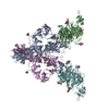
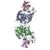
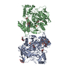
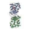
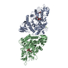
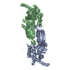
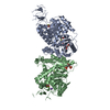
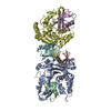
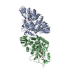
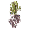
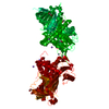
| Entry | Database: PDB / ID: 3nvn | |||||||||
|---|---|---|---|---|---|---|---|---|---|---|
| Title | Molecular mechanism of guidance cue recognition | |||||||||
 Components Components |
| |||||||||
 Keywords Keywords |  Viral Protein/SIGNALING PROTEIN / Beta-Propeller / signaling / Viral Protein-SIGNALING PROTEIN complex Viral Protein/SIGNALING PROTEIN / Beta-Propeller / signaling / Viral Protein-SIGNALING PROTEIN complex | |||||||||
| Function / homology |  Function and homology information Function and homology informationregulation of synapse pruning / semaphorin receptor binding / cerebellar climbing fiber to Purkinje cell synapse / Other semaphorin interactions / semaphorin-plexin signaling pathway involved in axon guidance / semaphorin receptor complex / semaphorin receptor activity / negative regulation of cell adhesion / positive regulation of axonogenesis / regulation of cell migration ...regulation of synapse pruning / semaphorin receptor binding / cerebellar climbing fiber to Purkinje cell synapse / Other semaphorin interactions / semaphorin-plexin signaling pathway involved in axon guidance / semaphorin receptor complex / semaphorin receptor activity / negative regulation of cell adhesion / positive regulation of axonogenesis / regulation of cell migration / regulation of cell shape / cell adhesion / signaling receptor binding / extracellular region / membrane / plasma membraneSimilarity search - Function | |||||||||
| Biological species |  Ectromelia virus Ectromelia virus Homo sapiens (human) Homo sapiens (human) | |||||||||
| Method | X-RAY DIFFRACTION / SYNCHROTRON / MOLECULAR REPLACEMENT / Resolution: 2.26 Å | |||||||||
 Authors Authors | Liu, H. / Juo, Z. / Shim, A. / Focia, P. / Chen, X. / Garcia, C. / He, X. | |||||||||
 Citation Citation | Journal: Cell(Cambridge,Mass.) / Year: 2010 Title: Structural Basis of Semaphorin-Plexin Recognition and Viral Mimicry from Sema7A and A39R Complexes with PlexinC1. Authors: Liu, H. / Juo, Z.S. / Shim, A.H. / Focia, P.J. / Chen, X. / Garcia, K.C. / He, X. | |||||||||
| History |
|
- Structure visualization
| Structure viewer | Molecule: MolmilJmol/JSmol |
|---|
- Downloads & links
-Download
| PDBx/mmCIF format | 3nvn.cif.gz | 192 KB | Display | PDBx/mmCIF format |
|---|---|---|---|---|
| PDB format | pdb3nvn.ent.gz | 153.5 KB | Display | PDB format |
| PDBx/mmJSON format | 3nvn.json.gz | Tree view | PDBx/mmJSON format | |
| Others |  Other downloads Other downloads |
-Validation report
| Arichive directory | https://data.pdbj.org/pub/pdb/validation_reports/nv/3nvnftp://data.pdbj.org/pub/pdb/validation_reports/nv/3nvn | HTTPS FTP |
|---|
-Related structure data

-Links
 PDBj
PDBj


- Assembly
| Deposited unit | 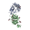
| ||||||||
|---|---|---|---|---|---|---|---|---|---|
| 1 |
| ||||||||
| Unit cell |
|
-Components
-Protein , 2 types, 2 molecules AB
| #1: Protein | Mass: 44037.844 Da / Num. of mol.: 1 / Fragment: UNP residues 15-399 Source method: isolated from a genetically manipulated source Source: (gene. exp.) Ectromelia virus / Gene: SEMA / Cell (production host): HEK293S / Production host: Homo sapiens (human) / References: UniProt: Q8JL80 |
|---|---|
| #2: Protein | Mass: 51800.102 Da / Num. of mol.: 1 / Fragment: UNP residues 35-507 Source method: isolated from a genetically manipulated source Source: (gene. exp.) Homo sapiens (human) / Gene: PLXNC1, VESPR / Cell (production host): HEK293S / Production host: Homo sapiens (human) / References: UniProt: O60486 |
-Sugars , 2 types, 8 molecules 

| #3: Sugar | N-Acetylglucosamine Type: D-saccharide, alpha linking / Mass: 221.208 Da / Num. of mol.: 3 Type: D-saccharide, alpha linking / Mass: 221.208 Da / Num. of mol.: 3Source method: isolated from a genetically manipulated source Formula: C8H15NO6 #5: Sugar | ChemComp-NAG / N-Acetylglucosamine Type: D-saccharide, beta linking / Mass: 221.208 Da / Num. of mol.: 5 Type: D-saccharide, beta linking / Mass: 221.208 Da / Num. of mol.: 5Source method: isolated from a genetically manipulated source Formula: C8H15NO6 |
|---|
-Non-polymers , 2 types, 510 molecules 

| #4: Chemical | ChemComp-CA /  Mass: 40.078 Da / Num. of mol.: 1 / Source method: obtained synthetically / Formula: Ca Mass: 40.078 Da / Num. of mol.: 1 / Source method: obtained synthetically / Formula: Ca |
|---|---|
| #6: Water | ChemComp-HOH / WaterMass: 18.015 Da / Num. of mol.: 509 / Source method: isolated from a natural source / Formula: H2O |
-Experimental details
-Experiment
| Experiment | Method: X-RAY DIFFRACTION / Number of used crystals: 1 |
|---|
- Sample preparation
| Crystal | Density Matthews: 2.85 Å3/Da / Density % sol: 56.79 % |
|---|---|
| Crystal grow | Temperature: 295 K / Method: vapor diffusion, sitting drop / pH: 7 Details: 6% PEG8000, 0.1M HEPES pH7.0, 0.2M NaCl, 0.1M CaCl2, VAPOR DIFFUSION, SITTING DROP, temperature 295K |
-Data collection
| Diffraction | Mean temperature: 100 K |
|---|---|
| Diffraction source | Source: SYNCHROTRON / Site: APS  / Beamline: 21-ID-G / Wavelength: 0.97856 Å / Beamline: 21-ID-G / Wavelength: 0.97856 Å |
| Detector | Type: MAR scanner 300 mm plate / Detector: IMAGE PLATE / Date: Feb 6, 2010 |
| Radiation | Protocol: SINGLE WAVELENGTH / Monochromatic (M) / Laue (L): M / Scattering type: x-ray |
| Radiation wavelength | Wavelength: 0.97856 Å / Relative weight: 1 |
| Reflection | Resolution: 2→50 Å / Num. obs: 73704 / Observed criterion σ(F): 0 / Observed criterion σ(I): 0 / Redundancy: 8.4 % / Biso Wilson estimate: 13.2 Å2 / Rmerge(I) obs: 0.088 / Net I/σ(I): 14.89 |
| Reflection shell | Resolution: 2→2.1 Å / Redundancy: 4 % / Rmerge(I) obs: 0.293 / Mean I/σ(I) obs: 2.97 / % possible all: 95.5 |
- Processing
| Software |
| ||||||||||||||||||||||||||||||||||||||||||||||||||||||||||||
|---|---|---|---|---|---|---|---|---|---|---|---|---|---|---|---|---|---|---|---|---|---|---|---|---|---|---|---|---|---|---|---|---|---|---|---|---|---|---|---|---|---|---|---|---|---|---|---|---|---|---|---|---|---|---|---|---|---|---|---|---|---|
| Refinement | Method to determine structure: MOLECULAR REPLACEMENT / Resolution: 2.26→43.53 Å / Rfactor Rfree error: 0.005 / Data cutoff high absF: 6766291.52 / Data cutoff low absF: 0 / Isotropic thermal model: RESTRAINED / Cross valid method: THROUGHOUT / σ(F): 0 / Stereochemistry target values: ENGH & HUBER / Details: BULK SOLVENT MODEL USED
| ||||||||||||||||||||||||||||||||||||||||||||||||||||||||||||
| Solvent computation | Solvent model: FLAT MODEL / Bsol: 39.78 Å2 / ksol: 0.31 e/Å3 | ||||||||||||||||||||||||||||||||||||||||||||||||||||||||||||
| Displacement parameters | Biso mean: 47.9 Å2
| ||||||||||||||||||||||||||||||||||||||||||||||||||||||||||||
| Refine analyze |
| ||||||||||||||||||||||||||||||||||||||||||||||||||||||||||||
| Refinement step | Cycle: LAST / Resolution: 2.26→43.53 Å
| ||||||||||||||||||||||||||||||||||||||||||||||||||||||||||||
| Refine LS restraints |
| ||||||||||||||||||||||||||||||||||||||||||||||||||||||||||||
| Refine LS restraints NCS | NCS model details: NONE | ||||||||||||||||||||||||||||||||||||||||||||||||||||||||||||
| LS refinement shell | Resolution: 2.26→2.4 Å / Rfactor Rfree error: 0.019 / Total num. of bins used: 6
| ||||||||||||||||||||||||||||||||||||||||||||||||||||||||||||
| Xplor file |
|
