[English] 日本語
 Yorodumi
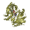
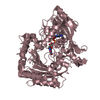
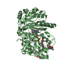
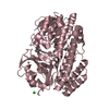
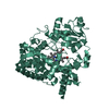
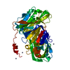
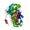
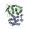
- PDB-6ml1: Structure of the USP15 deubiquitinase domain in complex with an a... -
+ Open data
- Basic information
| Entry | Database: PDB / ID: 6ml1 | ||||||
|---|---|---|---|---|---|---|---|
| Title | Structure of the USP15 deubiquitinase domain in complex with an affinity-matured inhibitory Ubv | ||||||
 Components Components |
| ||||||
 Keywords Keywords | SIGNALING PROTEIN / deubiuqitination / Ubv / high-affinity / inhibition | ||||||
| Function / homology |  Function and homology information Function and homology informationnegative regulation of antifungal innate immune response / regulation of intrinsic apoptotic signaling pathway in response to osmotic stress by p53 class mediator / protein K27-linked deubiquitination / positive regulation of RIG-I signaling pathway / regulation of RNA metabolic process / monoubiquitinated protein deubiquitination / ubiquitin-modified histone reader activity / transforming growth factor beta receptor binding / deubiquitinase activity / K48-linked deubiquitinase activity ...negative regulation of antifungal innate immune response / regulation of intrinsic apoptotic signaling pathway in response to osmotic stress by p53 class mediator / protein K27-linked deubiquitination / positive regulation of RIG-I signaling pathway / regulation of RNA metabolic process / monoubiquitinated protein deubiquitination / ubiquitin-modified histone reader activity / transforming growth factor beta receptor binding / deubiquitinase activity / K48-linked deubiquitinase activity / transcription elongation-coupled chromatin remodeling / SMAD binding / protein deubiquitination / BMP signaling pathway / transforming growth factor beta receptor signaling pathway / Downregulation of TGF-beta receptor signaling / negative regulation of transforming growth factor beta receptor signaling pathway / modification-dependent protein catabolic process / protein tag activity / UCH proteinases / ubiquitinyl hydrolase 1 / cysteine-type deubiquitinase activity / Ub-specific processing proteases / nuclear body / protein ubiquitination / cysteine-type endopeptidase activity / ubiquitin protein ligase binding / mitochondrion / proteolysis / nucleoplasm / metal ion binding / identical protein binding / nucleus / cytoplasm / cytosol Similarity search - Function | ||||||
| Biological species |  Homo sapiens (human) Homo sapiens (human) | ||||||
| Method |  X-RAY DIFFRACTION / X-RAY DIFFRACTION /  SYNCHROTRON / SYNCHROTRON /  MOLECULAR REPLACEMENT / Resolution: 1.9 Å MOLECULAR REPLACEMENT / Resolution: 1.9 Å | ||||||
 Authors Authors | Singer, A.U. / Teyra, J. / Boehmelt, G. / Lenter, M. / Sicheri, F. / Sidhu, S.S. | ||||||
 Citation Citation |  Journal: Structure / Year: 2019 Journal: Structure / Year: 2019Title: Structural and Functional Characterization of Ubiquitin Variant Inhibitors of USP15. Authors: Teyra, J. / Singer, A.U. / Schmitges, F.W. / Jaynes, P. / Kit Leng Lui, S. / Polyak, M.J. / Fodil, N. / Krieger, J.R. / Tong, J. / Schwerdtfeger, C. / Brasher, B.B. / Ceccarelli, D.F.J. / ...Authors: Teyra, J. / Singer, A.U. / Schmitges, F.W. / Jaynes, P. / Kit Leng Lui, S. / Polyak, M.J. / Fodil, N. / Krieger, J.R. / Tong, J. / Schwerdtfeger, C. / Brasher, B.B. / Ceccarelli, D.F.J. / Moffat, J. / Sicheri, F. / Moran, M.F. / Gros, P. / Eichhorn, P.J.A. / Lenter, M. / Boehmelt, G. / Sidhu, S.S. | ||||||
| History |
|
- Structure visualization
| Structure viewer | Molecule:  Molmil Molmil Jmol/JSmol Jmol/JSmol |
|---|
- Downloads & links
- Download
| PDBx/mmCIF format |  6ml1.cif.gz 6ml1.cif.gz | 190.6 KB | Display |  PDBx/mmCIF format PDBx/mmCIF format |
|---|---|---|---|---|
| PDB format |  pdb6ml1.ent.gz pdb6ml1.ent.gz | 146.3 KB | Display |  PDB format PDB format |
| PDBx/mmJSON format |  6ml1.json.gz 6ml1.json.gz | Tree view |  PDBx/mmJSON format PDBx/mmJSON format | |
| Others |  Other downloads Other downloads |
-Validation report
| Arichive directory |  https://data.pdbj.org/pub/pdb/validation_reports/ml/6ml1 https://data.pdbj.org/pub/pdb/validation_reports/ml/6ml1 ftp://data.pdbj.org/pub/pdb/validation_reports/ml/6ml1 ftp://data.pdbj.org/pub/pdb/validation_reports/ml/6ml1 | HTTPS FTP |
|---|
-Related structure data
| Related structure data |  6cpmC  6crnSC  6dj9C S: Starting model for refinement C: citing same article ( |
|---|---|
| Similar structure data |
- Links
Links
- Assembly
| Deposited unit | 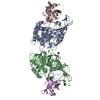
| ||||||||
|---|---|---|---|---|---|---|---|---|---|
| 1 | 
| ||||||||
| 2 | 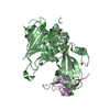
| ||||||||
| Unit cell |
|
- Components
-Protein , 2 types, 4 molecules ABEC
| #1: Protein | Mass: 39753.023 Da / Num. of mol.: 2 / Fragment: 275-470,781-934,275-470,781-934,275-470,781-934 Source method: isolated from a genetically manipulated source Source: (gene. exp.)  Homo sapiens (human) / Gene: USP15, KIAA0529 / Plasmid: pHH1013 Homo sapiens (human) / Gene: USP15, KIAA0529 / Plasmid: pHH1013Details (production host): N-terminal 6-His and Glutathione S-transferase (GST) tag followed by TEV cleavage site Production host:  #2: Protein | Mass: 10282.830 Da / Num. of mol.: 2 Source method: isolated from a genetically manipulated source Details: This is a Ubv (ubiquitin variant) selected by phage display to bindthe USP15 USP domain with high affinity Source: (gene. exp.)  Homo sapiens (human) / Gene: UBC / Plasmid: pET53 / Production host: Homo sapiens (human) / Gene: UBC / Plasmid: pET53 / Production host:  |
|---|
-Protein/peptide , 1 types, 1 molecules G
| #3: Protein/peptide | Mass: 3055.344 Da / Num. of mol.: 1 Source method: isolated from a genetically manipulated source Details: Typically this region is removed upon TEV cleavage. TEV cleavage was performed in this case, but I seem to have density which can only be attributed to residues from this proteolyzed N-terminal tag Source: (gene. exp.)  Details (production host): N-terminal region of the Ubv ending in a TEV cleavage site Production host:  |
|---|
-Non-polymers , 7 types, 402 molecules 


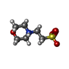


| #4: Chemical | ChemComp-CA / #5: Chemical | #6: Chemical | ChemComp-NA / #7: Chemical | ChemComp-EDO / | #8: Chemical | ChemComp-CL / | #9: Chemical | ChemComp-MES / | #10: Water | ChemComp-HOH / | |
|---|
-Experimental details
-Experiment
| Experiment | Method:  X-RAY DIFFRACTION / Number of used crystals: 1 X-RAY DIFFRACTION / Number of used crystals: 1 |
|---|
- Sample preparation
| Crystal | Density Matthews: 2.37 Å3/Da / Density % sol: 48.15 % |
|---|---|
| Crystal grow | Temperature: 295 K / Method: vapor diffusion, sitting drop / pH: 6.5 Details: 16% PEG3350, 100 mM MES 6.0, 250 mM CaCl2, cryoprotected with 18% PEG3350, 200 mM CaCl2, 100 mM MES pH 6.5 and 25% glycerol Temp details: Room temperature |
-Data collection
| Diffraction | Mean temperature: 100 K / Serial crystal experiment: N |
|---|---|
| Diffraction source | Source:  SYNCHROTRON / Site: SYNCHROTRON / Site:  APS APS  / Beamline: 24-ID-E / Wavelength: 0.97918 Å / Beamline: 24-ID-E / Wavelength: 0.97918 Å |
| Detector | Type: DECTRIS EIGER X 16M / Detector: PIXEL / Date: Apr 4, 2017 / Details: mirrors |
| Radiation | Protocol: SINGLE WAVELENGTH / Monochromatic (M) / Laue (L): M / Scattering type: x-ray |
| Radiation wavelength | Wavelength: 0.97918 Å / Relative weight: 1 |
| Reflection | Resolution: 1.9→95.27 Å / Num. obs: 71233 / % possible obs: 98.1 % / Redundancy: 3.9 % / CC1/2: 0.998 / Rmerge(I) obs: 0.031 / Net I/σ(I): 15.5 |
| Reflection shell | Resolution: 1.9→1.94 Å / Redundancy: 3.2 % / Rmerge(I) obs: 0.317 / Mean I/σ(I) obs: 2.9 / Num. unique obs: 3864 / CC1/2: 0.987 / % possible all: 81.6 |
- Processing
| Software |
| |||||||||||||||||||||||||||||||||||||||||||||||||||||||||||||||||||||||||||||||||||||||||||||||||||||||||||||||||||||||||||||||||||||||||||||||||||||||||||||||||||||||||||||||
|---|---|---|---|---|---|---|---|---|---|---|---|---|---|---|---|---|---|---|---|---|---|---|---|---|---|---|---|---|---|---|---|---|---|---|---|---|---|---|---|---|---|---|---|---|---|---|---|---|---|---|---|---|---|---|---|---|---|---|---|---|---|---|---|---|---|---|---|---|---|---|---|---|---|---|---|---|---|---|---|---|---|---|---|---|---|---|---|---|---|---|---|---|---|---|---|---|---|---|---|---|---|---|---|---|---|---|---|---|---|---|---|---|---|---|---|---|---|---|---|---|---|---|---|---|---|---|---|---|---|---|---|---|---|---|---|---|---|---|---|---|---|---|---|---|---|---|---|---|---|---|---|---|---|---|---|---|---|---|---|---|---|---|---|---|---|---|---|---|---|---|---|---|---|---|---|---|
| Refinement | Method to determine structure:  MOLECULAR REPLACEMENT MOLECULAR REPLACEMENTStarting model: USP15 and Ubv from 6CRN Resolution: 1.9→49.319 Å / SU ML: 0.21 / Cross valid method: FREE R-VALUE / σ(F): 1.34 / Phase error: 23.13
| |||||||||||||||||||||||||||||||||||||||||||||||||||||||||||||||||||||||||||||||||||||||||||||||||||||||||||||||||||||||||||||||||||||||||||||||||||||||||||||||||||||||||||||||
| Solvent computation | Shrinkage radii: 0.9 Å / VDW probe radii: 1.11 Å | |||||||||||||||||||||||||||||||||||||||||||||||||||||||||||||||||||||||||||||||||||||||||||||||||||||||||||||||||||||||||||||||||||||||||||||||||||||||||||||||||||||||||||||||
| Refinement step | Cycle: LAST / Resolution: 1.9→49.319 Å
| |||||||||||||||||||||||||||||||||||||||||||||||||||||||||||||||||||||||||||||||||||||||||||||||||||||||||||||||||||||||||||||||||||||||||||||||||||||||||||||||||||||||||||||||
| Refine LS restraints |
| |||||||||||||||||||||||||||||||||||||||||||||||||||||||||||||||||||||||||||||||||||||||||||||||||||||||||||||||||||||||||||||||||||||||||||||||||||||||||||||||||||||||||||||||
| LS refinement shell |
|
 Movie
Movie Controller


 PDBj
PDBj


