Resolution: 2.28→48.057 Å / Num. obs: 105055 / % possible obs: 94.6 % / Observed criterion σ(I): -3 / Redundancy: 2.75 % / Biso Wilson estimate: 41.55 Å2 / Rmerge(I) obs: 0.054 / Net I/σ(I): 13.25
Reflection shell
Diffraction-ID: 1
Resolution (Å)
Redundancy (%)
Rmerge(I) obs
Mean I/σ(I) obs
Num. measured obs
Num. unique obs
% possible all
2.28-2.36
2.09
0.446
2.1
15353
7352
67.6
2.36-2.46
0.407
2.5
25143
10783
91.9
2.46-2.57
0.355
3.2
30301
10770
99
2.57-2.7
0.284
4.1
30176
10516
98.6
2.7-2.87
0.197
5.8
31896
11092
98.9
2.87-3.09
0.125
8.7
31556
10941
98.5
3.09-3.4
0.074
13.8
31402
10912
98.3
3.4-3.89
0.039
22.8
31394
10888
97.8
3.89-4.89
0.026
30.9
31005
10815
97.3
4.89-48.057
0.022
34.5
31089
10985
97.6
-
Phasing
Phasing
Method: MAD
-
Processing
Software
Name
Version
Classification
NB
REFMAC
5.4.0067
refinement
PHENIX
refinement
SHELX
phasing
MolProbity
3beta29
modelbuilding
XSCALE
datascaling
PDB_EXTRACT
3
dataextraction
ADSC
Quantum
datacollection
XDS
datareduction
SHELXD
phasing
autoSHARP
phasing
Refinement
Method to determine structure: MAD / Resolution: 2.28→48.057 Å / Cor.coef. Fo:Fc: 0.963 / Cor.coef. Fo:Fc free: 0.942 / SU B: 13.379 / SU ML: 0.162 / TLS residual ADP flag: LIKELY RESIDUAL / Cross valid method: THROUGHOUT / σ(F): 0 / ESU R: 0.26 / ESU R Free: 0.204 Stereochemistry target values: MAXIMUM LIKELIHOOD WITH PHASES Details: 1. HYDROGENS HAVE BEEN ADDED IN THE RIDING POSITIONS. 2. A MET-INHIBITION PROTOCOL WAS USED FOR SELENOMETHIONINE INCORPORATION DURING PROTEIN EXPRESSION. THE OCCUPANCY OF THE SE ATOMS IN THE ...Details: 1. HYDROGENS HAVE BEEN ADDED IN THE RIDING POSITIONS. 2. A MET-INHIBITION PROTOCOL WAS USED FOR SELENOMETHIONINE INCORPORATION DURING PROTEIN EXPRESSION. THE OCCUPANCY OF THE SE ATOMS IN THE MSE RESIDUES WAS REDUCED TO 0.75 FOR THE REDUCED SCATTERING POWER DUE TO PARTIAL S-MET INCORPORATION. 3. ATOM RECORD CONTAINS RESIDUAL B FACTORS ONLY. 4. EDO AND CL WERE MODELED BASED ON CRYSTALLIZATION CONDITIONS. MG WAS TENTATIVELY MODELED BASED ON ENVIRONMENT.
Rfactor
Num. reflection
% reflection
Selection details
Rfree
0.223
5271
5 %
RANDOM
Rwork
0.18
-
-
-
obs
0.182
105052
95.14 %
-
Solvent computation
Ion probe radii: 0.8 Å / Shrinkage radii: 0.8 Å / VDW probe radii: 1.2 Å / Solvent model: MASK
Displacement parameters
Biso mean: 40.479 Å2
Baniso -1
Baniso -2
Baniso -3
1-
-0.86 Å2
-0.43 Å2
0 Å2
2-
-
-0.86 Å2
0 Å2
3-
-
-
1.3 Å2
Refinement step
Cycle: LAST / Resolution: 2.28→48.057 Å
Protein
Nucleic acid
Ligand
Solvent
Total
Num. atoms
14264
0
125
540
14929
Refine LS restraints
Refine-ID
Type
Dev ideal
Dev ideal target
Number
X-RAY DIFFRACTION
r_bond_refined_d
0.013
0.021
14699
X-RAY DIFFRACTION
r_bond_other_d
0.002
0.02
9897
X-RAY DIFFRACTION
r_angle_refined_deg
1.478
1.944
19835
X-RAY DIFFRACTION
r_angle_other_deg
1.571
3
24036
X-RAY DIFFRACTION
r_dihedral_angle_1_deg
6.633
5
1839
X-RAY DIFFRACTION
r_dihedral_angle_2_deg
33.297
24.052
686
X-RAY DIFFRACTION
r_dihedral_angle_3_deg
15.725
15
2362
X-RAY DIFFRACTION
r_dihedral_angle_4_deg
20.225
15
74
X-RAY DIFFRACTION
r_chiral_restr
0.074
0.2
2154
X-RAY DIFFRACTION
r_gen_planes_refined
0.006
0.02
16533
X-RAY DIFFRACTION
r_gen_planes_other
0.003
0.02
3073
X-RAY DIFFRACTION
r_mcbond_it
1.262
3
9112
X-RAY DIFFRACTION
r_mcbond_other
0.435
3
3787
X-RAY DIFFRACTION
r_mcangle_it
2.386
5
14612
X-RAY DIFFRACTION
r_scbond_it
4.305
8
5587
X-RAY DIFFRACTION
r_scangle_it
6.434
11
5218
Refine LS restraints NCS
Ens-ID: 1 / Refine-ID: X-RAY DIFFRACTION
Dom-ID
Auth asym-ID
Number
Type
Rms dev position (Å)
Weight position
1
A
2523
TIGHTPOSITIONAL
0.21
0.2
2
B
2523
TIGHTPOSITIONAL
0.18
0.2
3
C
2523
TIGHTPOSITIONAL
0.14
0.2
4
D
2523
TIGHTPOSITIONAL
0.13
0.2
1
A
2940
MEDIUMPOSITIONAL
0.28
0.5
2
B
2940
MEDIUMPOSITIONAL
0.34
0.5
3
C
2940
MEDIUMPOSITIONAL
0.23
0.5
4
D
2940
MEDIUMPOSITIONAL
0.23
0.5
1
A
181
LOOSEPOSITIONAL
0.86
5
2
B
181
LOOSEPOSITIONAL
0.68
5
3
C
181
LOOSEPOSITIONAL
0.7
5
4
D
181
LOOSEPOSITIONAL
0.63
5
1
A
2523
TIGHTTHERMAL
0.81
2
2
B
2523
TIGHTTHERMAL
0.73
2
3
C
2523
TIGHTTHERMAL
0.6
2
4
D
2523
TIGHTTHERMAL
0.66
2
1
A
2940
MEDIUMTHERMAL
0.96
4
2
B
2940
MEDIUMTHERMAL
0.86
4
3
C
2940
MEDIUMTHERMAL
0.73
4
4
D
2940
MEDIUMTHERMAL
0.79
4
1
A
181
LOOSETHERMAL
2.23
10
2
B
181
LOOSETHERMAL
1.99
10
3
C
181
LOOSETHERMAL
1.56
10
4
D
181
LOOSETHERMAL
1.71
10
LS refinement shell
Resolution: 2.28→2.344 Å / Total num. of bins used: 20
Rfactor
Num. reflection
% reflection
Rfree
0.324
282
-
Rwork
0.265
5367
-
all
-
5649
-
obs
-
-
69.54 %
Refinement TLS params.
Method: refined / Refine-ID: X-RAY DIFFRACTION
ID
L11 (°2)
L12 (°2)
L13 (°2)
L22 (°2)
L23 (°2)
L33 (°2)
S11 (Å °)
S12 (Å °)
S13 (Å °)
S21 (Å °)
S22 (Å °)
S23 (Å °)
S31 (Å °)
S32 (Å °)
S33 (Å °)
T11 (Å2)
T12 (Å2)
T13 (Å2)
T22 (Å2)
T23 (Å2)
T33 (Å2)
Origin x (Å)
Origin y (Å)
Origin z (Å)
1
1.2767
-0.1324
-0.5961
0.4169
0.0357
1.213
-0.1608
0.1501
-0.2793
-0.1101
0.0903
0.0563
0.1672
-0.1965
0.0705
-0.2578
-0.0501
0.0326
-0.1632
0.0485
-0.1446
147.5858
138.2775
66.2043
2
3.0673
1.1328
-0.6122
1.1477
-0.2915
0.884
-0.0852
0.4086
-0.0572
0.0696
-0.0063
-0.0717
0.1497
-0.0377
0.0915
-0.2296
-0.1141
0.0943
0.0326
0.03
-0.1155
105.9226
113.9186
90.5057
3
1.9546
0.357
0.5071
1.3352
0.15
1.8422
0.1678
-0.2828
0.0239
0.625
-0.1856
0.543
0.2043
-0.3392
0.0178
0.2138
-0.0918
0.355
0.0897
-0.0213
0.023
115.2932
143.2927
126.2309
4
0.9962
0.2478
0.2271
1.4187
-0.0724
1.3768
0.0187
-0.0347
0.4877
0.1895
0.0144
0.234
-0.4865
-0.2498
-0.033
-0.11
0.1468
0.0602
-0.0933
0.0794
0.0465
135.0474
175.1531
86.7762
Refinement TLS group
ID
Refine-ID
Refine TLS-ID
Auth asym-ID
Label asym-ID
Auth seq-ID
Label seq-ID
1
X-RAY DIFFRACTION
1
A
A
0 - 475
19 - 494
2
X-RAY DIFFRACTION
2
B
B
0 - 475
19 - 494
3
X-RAY DIFFRACTION
3
C
C
0 - 475
19 - 494
4
X-RAY DIFFRACTION
4
D
D
0 - 475
19 - 494
+
About Yorodumi
-
News
-
Feb 9, 2022. New format data for meta-information of EMDB entries
New format data for meta-information of EMDB entries
Version 3 of the EMDB header file is now the official format.
The previous official version 1.9 will be removed from the archive.
In the structure databanks used in Yorodumi, some data are registered as the other names, "COVID-19 virus" and "2019-nCoV". Here are the details of the virus and the list of structure data.
Jan 31, 2019. EMDB accession codes are about to change! (news from PDBe EMDB page)
EMDB accession codes are about to change! (news from PDBe EMDB page)
The allocation of 4 digits for EMDB accession codes will soon come to an end. Whilst these codes will remain in use, new EMDB accession codes will include an additional digit and will expand incrementally as the available range of codes is exhausted. The current 4-digit format prefixed with “EMD-” (i.e. EMD-XXXX) will advance to a 5-digit format (i.e. EMD-XXXXX), and so on. It is currently estimated that the 4-digit codes will be depleted around Spring 2019, at which point the 5-digit format will come into force.
The EM Navigator/Yorodumi systems omit the EMD- prefix.
Related info.:Q: What is EMD? / ID/Accession-code notation in Yorodumi/EM Navigator
Yorodumi is a browser for structure data from EMDB, PDB, SASBDB, etc.
This page is also the successor to EM Navigator detail page, and also detail information page/front-end page for Omokage search.
The word "yorodu" (or yorozu) is an old Japanese word meaning "ten thousand". "mi" (miru) is to see.
Related info.:EMDB / PDB / SASBDB / Comparison of 3 databanks / Yorodumi Search / Aug 31, 2016. New EM Navigator & Yorodumi / Yorodumi Papers / Jmol/JSmol / Function and homology information / Changes in new EM Navigator and Yorodumi
 Movie
Movie Controller

 Yorodumi
 Open data
 Basic information
 Components
 Keywords
 Function and homology information
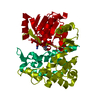
 Desulfovibrio desulfuricans subsp. desulfuricans str. (bacteria)
 X-RAY DIFFRACTION /
  Authors
 Citation
 Structure visualization
 Downloads & links
 Other downloads
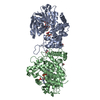


 PDBj
PDBj


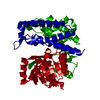
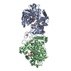
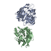
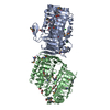
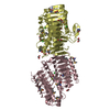
 Assembly

 Mass: 35.453 Da / Num. of mol.: 12 / Source method: obtained synthetically / Formula: Cl
 Mass: 62.068 Da / Num. of mol.: 28 / Source method: obtained synthetically / Formula: C2H6O2
 Mass: 24.305 Da / Num. of mol.: 1 / Source method: obtained synthetically / Formula: Mg
 Mass: 18.015 Da / Num. of mol.: 540 / Source method: isolated from a natural source / Formula: H2O
 Sample preparation
 / Beamline: 8.2.1 / Wavelength: 1.0000, 0.9795, 0.9797
 Processing