 Movie
Movie Controller

[English] 日本語
 Yorodumi
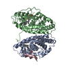
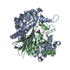
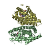
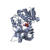
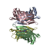
- PDB-1nfp: STRUCTURAL REFINEMENT OF THE NON-FLUORESCENT FLAVOPROTEIN FROM PH... -
+ Open data

- Basic information
| Entry | Database: PDB / ID: 1nfp | ||||||
|---|---|---|---|---|---|---|---|
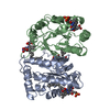
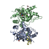
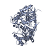
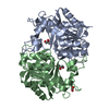
| Title | STRUCTURAL REFINEMENT OF THE NON-FLUORESCENT FLAVOPROTEIN FROM PHOTOBACTERIUM LEIOGNATHI AT 1.60 ANGSTROMS RESOLUTION | ||||||
 Components Components | LUXF GENE PRODUCT | ||||||
 Keywords Keywords |  FLAVOPROTEIN / FLAVIN MONONUCLEOTIDE / MYRISTATE FLAVOPROTEIN / FLAVIN MONONUCLEOTIDE / MYRISTATE | ||||||
| Function / homology |  Function and homology information Function and homology information | ||||||
| Biological species |  Photobacterium leiognathi (bacteria) Photobacterium leiognathi (bacteria) | ||||||
| Method | X-RAY DIFFRACTION / SYNCHROTRON / Resolution: 1.6 Å | ||||||
 Authors Authors | Moore, S.A. / Njames, M.N.G. | ||||||
 Citation Citation | Journal: J.Mol.Biol. / Year: 1995 Title: Structural refinement of the non-fluorescent flavoprotein from Photobacterium leiognathi at 1.60 A resolution. Authors: Moore, S.A. / James, M.N. #1: Journal: Protein Sci. / Year: 1994Title: Common Structural Features of the Luxf Protein and the Subunits of Bacterial Luciferase: Evidence for a (Beta(Slash)Alpha)8 Fold in Luciferase Authors: Moore, S.A. / James, M.N.G. #2: Journal: Embo J. / Year: 1993Title: Crystal Structure of a Flavoprotein Related to the Subunits of Bacterial Luciferase Authors: Moore, S.A. / James, M.N.G. / O'Kane, D.J. / Lee, J. #3: Journal: J.Mol.Biol. / Year: 1992Title: Crystallization of Photobacterium Leiognathi Nfp, Non-Fluorescent Flavoprotein an Unusual Flavoprotein with Limited Sequence Identity to Bacterial Luciferase Authors: Moore, S.A. / James, M.N.G. / O'Kane, D.J. / Lee, J. | ||||||
| History |
|
- Structure visualization
| Structure viewer | Molecule: MolmilJmol/JSmol |
|---|
- Downloads & links
-Download
| PDBx/mmCIF format | 1nfp.cif.gz | 67.3 KB | Display | PDBx/mmCIF format |
|---|---|---|---|---|
| PDB format | pdb1nfp.ent.gz | 49 KB | Display | PDB format |
| PDBx/mmJSON format | 1nfp.json.gz | Tree view | PDBx/mmJSON format | |
| Others |  Other downloads Other downloads |
-Validation report
| Arichive directory | https://data.pdbj.org/pub/pdb/validation_reports/nf/1nfpftp://data.pdbj.org/pub/pdb/validation_reports/nf/1nfp | HTTPS FTP |
|---|
-Related structure data
| Similar structure data |
|---|

-Links
 PDBj
PDBj


- Assembly
| Deposited unit | 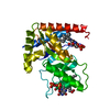
| |||||||||
|---|---|---|---|---|---|---|---|---|---|---|
| 1 | 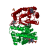
| |||||||||
| Unit cell |
| |||||||||
| Components on special symmetry positions |
| |||||||||
| Details | SYMMETRY THE CRYSTALLOGRAPHIC SYMMETRY TRANSFORMATIONS PRESENTED BELOW GENERATE THE SUBUNITS OF THE POLYMERIC MOLECULE. APPLIED TO RESIDUES: 1 1 .. 423 TWO-FOLD AXIS AT 0,Y,1/4: THE MOLECULE IS A SYMMETRICAL HOMODIMER. SYMMETRY1 1 -1.000000 0.000000 0.000000 0.00000 SYMMETRY2 1 0.000000 1.000000 0.000000 0.00000 SYMMETRY3 1 0.000000 0.000000 -1.000000 49.66500 |
-Components
| #1: Protein | Mass: 26312.686 Da / Num. of mol.: 1 Source method: isolated from a genetically manipulated source Source: (gene. exp.) Photobacterium leiognathi (bacteria) / References: UniProt: P09142 | ||||||
|---|---|---|---|---|---|---|---|
| #2: Chemical | ChemComp-SO4 / Sulfate  Mass: 96.063 Da / Num. of mol.: 1 / Source method: obtained synthetically / Formula: SO4 Mass: 96.063 Da / Num. of mol.: 1 / Source method: obtained synthetically / Formula: SO4 | ||||||
| #3: Chemical | Flavin mononucleotide  Mass: 456.344 Da / Num. of mol.: 2 / Source method: obtained synthetically / Formula: C17H21N4O9P Mass: 456.344 Da / Num. of mol.: 2 / Source method: obtained synthetically / Formula: C17H21N4O9P#4: Chemical | Myristic acid  Mass: 228.371 Da / Num. of mol.: 2 / Source method: obtained synthetically / Formula: C14H28O2 Mass: 228.371 Da / Num. of mol.: 2 / Source method: obtained synthetically / Formula: C14H28O2#5: Water | ChemComp-HOH / | Water Mass: 18.015 Da / Num. of mol.: 192 / Source method: isolated from a natural source / Formula: H2O Mass: 18.015 Da / Num. of mol.: 192 / Source method: isolated from a natural source / Formula: H2OCompound details | THE STRUCTURAL FORMULA HYDROGEN ATOMS EQUIVALENTS PERTAIN TO THE OXIDIZED STATE OF FMN. THE ...THE STRUCTURAL | |
-Experimental details
-Experiment
| Experiment | Method: X-RAY DIFFRACTION / Number of used crystals: 2 |
|---|
- Sample preparation
| Crystal | Density Matthews: 2.47 Å3/Da / Density % sol: 50.3 % / Description: DATA COLLECTED FROM TWO CRYSTALS | |||||||||||||||||||||||||
|---|---|---|---|---|---|---|---|---|---|---|---|---|---|---|---|---|---|---|---|---|---|---|---|---|---|---|
| Crystal | *PLUS Density % sol: 51 % | |||||||||||||||||||||||||
| Crystal grow | *PLUS pH: 5.5 / Method: vapor diffusion, hanging drop | |||||||||||||||||||||||||
| Components of the solutions | *PLUS
|
-Data collection
| Diffraction source | Source: SYNCHROTRON / Site: Photon Factory  / Beamline: BL-6A / Wavelength: 1 Å / Beamline: BL-6A / Wavelength: 1 Å |
|---|---|
| Detector | Type: FUJI / Detector: IMAGE PLATE / Date: Nov 1, 1992 |
| Radiation | Scattering type: x-ray |
| Radiation wavelength | Wavelength: 1 Å / Relative weight: 1 |
| Reflection | Num. obs: 28223 / % possible obs: 81 % / Observed criterion σ(I): 0 / Redundancy: 3 % / Rmerge(I) obs: 0.059 |
| Reflection | *PLUS Highest resolution: 1.6 Å / Lowest resolution: 9999 Å / Num. measured all: 84138 / Rmerge(I) obs: 0.059 / Biso Wilson estimate: 16.07 Å2 |
| Reflection shell | *PLUS Highest resolution: 1.6 Å / Lowest resolution: 1.67 Å / % possible obs: 65.6 % / Num. unique obs: 2590 / Num. measured obs: 3954 |
- Processing
| Software |
| ||||||||||||||||||||||||||||||||||||||||||||||||||||||||||||||||||||||||||||||||||||
|---|---|---|---|---|---|---|---|---|---|---|---|---|---|---|---|---|---|---|---|---|---|---|---|---|---|---|---|---|---|---|---|---|---|---|---|---|---|---|---|---|---|---|---|---|---|---|---|---|---|---|---|---|---|---|---|---|---|---|---|---|---|---|---|---|---|---|---|---|---|---|---|---|---|---|---|---|---|---|---|---|---|---|---|---|---|
| Refinement | Resolution: 1.6→10 Å / σ(F): 1 /
| ||||||||||||||||||||||||||||||||||||||||||||||||||||||||||||||||||||||||||||||||||||
| Refinement step | Cycle: LAST / Resolution: 1.6→10 Å
| ||||||||||||||||||||||||||||||||||||||||||||||||||||||||||||||||||||||||||||||||||||
| Refine LS restraints |
|
