 Movie
Movie Controller

[English] 日本語
 Yorodumi
- PDB-1dug: STRUCTURE OF THE FIBRINOGEN G CHAIN INTEGRIN BINDING AND FACTOR X... -
+ Open data

- Basic information
| Entry | Database: PDB / ID: 1dug | ||||||
|---|---|---|---|---|---|---|---|
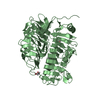
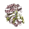
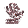
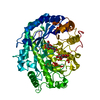
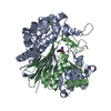
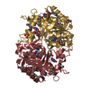
| Title | STRUCTURE OF THE FIBRINOGEN G CHAIN INTEGRIN BINDING AND FACTOR XIIIA CROSSLINKING SITES OBTAINED THROUGH CARRIER PROTEIN DRIVEN CRYSTALLIZATION | ||||||
 Components Components | chimera of GLUTATHIONE S-TRANSFERASE-synthetic LINKEr-C-TERMINAL FIBRINOGEN GAMMA CHAIN | ||||||
 Keywords Keywords |  transferase / blood clotting / gamma chain integrin fragment / carrier protein driven crystallization transferase / blood clotting / gamma chain integrin fragment / carrier protein driven crystallization | ||||||
| Function / homology |  Function and homology information Function and homology informationplatelet maturation / fibrinogen complex / Regulation of TLR by endogenous ligand / blood coagulation, fibrin clot formation / platelet alpha granule / cellular response to interleukin-6 / MyD88 deficiency (TLR2/4) / positive regulation of heterotypic cell-cell adhesion / IRAK4 deficiency (TLR2/4) / MyD88:MAL(TIRAP) cascade initiated on plasma membrane ...platelet maturation / fibrinogen complex / Regulation of TLR by endogenous ligand / blood coagulation, fibrin clot formation / platelet alpha granule / cellular response to interleukin-6 / MyD88 deficiency (TLR2/4) / positive regulation of heterotypic cell-cell adhesion / IRAK4 deficiency (TLR2/4) / MyD88:MAL(TIRAP) cascade initiated on plasma membrane / extracellular matrix structural constituent / plasminogen activation / p130Cas linkage to MAPK signaling for integrins / positive regulation of peptide hormone secretion / positive regulation of exocytosis / GRB2:SOS provides linkage to MAPK signaling for Integrins / glutathione transferase / glutathione transferase activity / protein secretion / protein polymerization / Integrin cell surface interactions / cellular response to interleukin-1 / negative regulation of endothelial cell apoptotic process / Common Pathway of Fibrin Clot Formation / positive regulation of substrate adhesion-dependent cell spreading / negative regulation of extrinsic apoptotic signaling pathway via death domain receptors / positive regulation of vasoconstriction / fibrinolysis / cell adhesion molecule binding / Integrin signaling / cell-matrix adhesion / platelet alpha granule lumen / Post-translational protein phosphorylation / positive regulation of protein secretion / Signaling by high-kinase activity BRAF mutants / MAP2K and MAPK activation / platelet aggregation / response to calcium ion / Regulation of Insulin-like Growth Factor (IGF) transport and uptake by Insulin-like Growth Factor Binding Proteins (IGFBPs) / Signaling by RAF1 mutants / Signaling by moderate kinase activity BRAF mutants / Paradoxical activation of RAF signaling by kinase inactive BRAF / Signaling downstream of RAS mutants / Signaling by BRAF and RAF1 fusions / Platelet degranulation / ER-Phagosome pathway / protein-containing complex assembly / collagen-containing extracellular matrix / blood microparticle / positive regulation of ERK1 and ERK2 cascade / endoplasmic reticulum lumen / external side of plasma membrane / signaling receptor binding / structural molecule activity / cell surface / extracellular space / extracellular exosome / extracellular region / identical protein binding / metal ion binding / plasma membraneSimilarity search - Function | ||||||
| Biological species |  Schistosoma japonicum (invertebrata) Schistosoma japonicum (invertebrata) Homo sapiens (human) Homo sapiens (human) | ||||||
| Method | X-RAY DIFFRACTION / SYNCHROTRON / Resolution: 1.8 Å | ||||||
 Authors Authors | Ware, S. / Donahue, J.P. / Hawiger, J. / Anderson, W.F. | ||||||
 Citation Citation | Journal: Protein Sci. / Year: 1999 Title: Structure of the fibrinogen gamma-chain integrin binding and factor XIIIa cross-linking sites obtained through carrier protein driven crystallization. Authors: Ware, S. / Donahue, J.P. / Hawiger, J. / Anderson, W.F. | ||||||
| History |
|
- Structure visualization
| Structure viewer | Molecule: MolmilJmol/JSmol |
|---|
- Downloads & links
-Download
| PDBx/mmCIF format | 1dug.cif.gz | 120.7 KB | Display | PDBx/mmCIF format |
|---|---|---|---|---|
| PDB format | pdb1dug.ent.gz | 94.5 KB | Display | PDB format |
| PDBx/mmJSON format | 1dug.json.gz | Tree view | PDBx/mmJSON format | |
| Others |  Other downloads Other downloads |
-Validation report
| Arichive directory | https://data.pdbj.org/pub/pdb/validation_reports/du/1dugftp://data.pdbj.org/pub/pdb/validation_reports/du/1dug | HTTPS FTP |
|---|
-Related structure data
| Similar structure data |
|---|
-Links
 PDBj
PDBj


- Assembly
| Deposited unit | 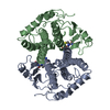
| ||||||||
|---|---|---|---|---|---|---|---|---|---|
| 1 |
| ||||||||
| Unit cell |
|
-Components
| #1: Protein | Mass: 27133.359 Da / Num. of mol.: 2 Source method: isolated from a genetically manipulated source Details: GLUTATHIONE S-TRANSFERASE (residues 1-217) bound to synthetic SDP linker (residues 218-220) bound to C-TERMINAL FIBRINOGEN GAMMA CHAIN (residues 221-234) Source: (gene. exp.) Schistosoma japonicum (invertebrata), (gene. exp.) Homo sapiens (human)Description: EUKARYOTA; METAZOA; PLATYHELMINTHES; TREMATODA; DIGENEA; STRIGEIDIDA; SCHISTOSO MATOIDEA; SCHISTOSOMATIDAE; SCHISTOSOMA Gene: FGG,PRO2061 / Production host:  Escherichia coli (E. coli) / Strain (production host): DH5ALPHA Escherichia coli (E. coli) / Strain (production host): DH5ALPHAReferences: UniProt: P08515, UniProt: P02679, glutathione transferase#2: Chemical | Glutathione  Mass: 307.323 Da / Num. of mol.: 2 / Source method: obtained synthetically / Formula: C10H17N3O6S Mass: 307.323 Da / Num. of mol.: 2 / Source method: obtained synthetically / Formula: C10H17N3O6S#3: Water | ChemComp-HOH / | Water Mass: 18.015 Da / Num. of mol.: 662 / Source method: isolated from a natural source / Formula: H2O Mass: 18.015 Da / Num. of mol.: 662 / Source method: isolated from a natural source / Formula: H2O |
|---|
-Experimental details
-Experiment
| Experiment | Method: X-RAY DIFFRACTION / Number of used crystals: 1 |
|---|
- Sample preparation
| Crystal | Density Matthews: 3.54 Å3/Da / Density % sol: 65.21 % | |||||||||||||||||||||||||||||||||||||||||||||||||||||||
|---|---|---|---|---|---|---|---|---|---|---|---|---|---|---|---|---|---|---|---|---|---|---|---|---|---|---|---|---|---|---|---|---|---|---|---|---|---|---|---|---|---|---|---|---|---|---|---|---|---|---|---|---|---|---|---|---|
| Crystal grow | Temperature: 277 K / Method: vapor diffusion, hanging drop / pH: 4.6 Details: PEG 3350, sodium acetate, sodium chloride, ammonium sulfate, Tris, reduced glutathione, pH 4.6, VAPOR DIFFUSION, HANGING DROP | |||||||||||||||||||||||||||||||||||||||||||||||||||||||
| Crystal grow | *PLUS Temperature: -170 ℃ | |||||||||||||||||||||||||||||||||||||||||||||||||||||||
| Components of the solutions | *PLUS
|
-Data collection
| Diffraction | Mean temperature: 100 K |
|---|---|
| Diffraction source | Source: SYNCHROTRON / Site: NSLS  / Beamline: X4A / Wavelength: 1.0093 / Beamline: X4A / Wavelength: 1.0093 |
| Detector | Type: FUJI / Detector: IMAGE PLATE / Date: Mar 15, 1997 |
| Radiation | Protocol: SINGLE WAVELENGTH / Monochromatic (M) / Laue (L): M / Scattering type: x-ray |
| Radiation wavelength | Wavelength: 1.0093 Å / Relative weight: 1 |
| Reflection | Resolution: 1.7→28 Å / Num. all: 376772 / Num. obs: 67736 / % possible obs: 89.2 % / Observed criterion σ(F): 0 / Observed criterion σ(I): -3 / Redundancy: 5.56 % / Rmerge(I) obs: 0.054 |
| Reflection shell | Resolution: 1.8→2 Å / Redundancy: 3.1 % / Rmerge(I) obs: 0.25 / Num. unique all: 12570 / % possible all: 66.1 |
| Reflection | *PLUS Num. measured all: 376772 |
| Reflection shell | *PLUS % possible obs: 66.1 % |
- Processing
| Software |
| |||||||||||||||||||||||||
|---|---|---|---|---|---|---|---|---|---|---|---|---|---|---|---|---|---|---|---|---|---|---|---|---|---|---|
| Refinement | Resolution: 1.8→28 Å / σ(F): 2 / σ(I): 0 / Stereochemistry target values: Engh & Huber
| |||||||||||||||||||||||||
| Refinement step | Cycle: LAST / Resolution: 1.8→28 Å
| |||||||||||||||||||||||||
| Refine LS restraints |
|
