 ムービー
ムービー コントローラー

+ データを開く

- 基本情報
基本情報
| 登録情報 | データベース: SASBDB / ID: SASDAS6 |
|---|---|
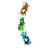
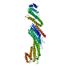
 試料 試料 | Plectin, fragment of the plakin domain encompassing the spectrin repeats SR3-SR4-SR5 and the SH3
|
| 機能・相同性 |  機能・相同性情報 機能・相同性情報protein-containing complex organization / actomyosin contractile ring assembly actin filament organization / tight junction organization / Type I hemidesmosome assembly / skeletal myofibril assembly / hemidesmosome assembly / hemidesmosome / leukocyte migration involved in immune response / intermediate filament organization / intermediate filament cytoskeleton organization ...protein-containing complex organization / actomyosin contractile ring assembly actin filament organization / tight junction organization / Type I hemidesmosome assembly / skeletal myofibril assembly / hemidesmosome assembly / hemidesmosome / leukocyte migration involved in immune response / intermediate filament organization / intermediate filament cytoskeleton organization / dystroglycan binding / fibroblast migration / cellular response to hydrostatic pressure / adherens junction organization / regulation of vascular permeability / costamere / T cell chemotaxis / cellular response to fluid shear stress / intermediate filament cytoskeleton / peripheral nervous system myelin maintenance / myoblast differentiation / cardiac muscle cell development / podosome / structural constituent of muscle / ankyrin binding / sarcomere organization / Assembly of collagen fibrils and other multimeric structures / response to food / nucleus organization / keratinocyte development / transmission of nerve impulse / brush border / sarcoplasm / Caspase-mediated cleavage of cytoskeletal proteins / establishment of skin barrier / skeletal muscle fiber development / respiratory electron transport chain / mitochondrion organization / wound healing / cellular response to mechanical stimulus / sarcolemma / structural constituent of cytoskeleton / multicellular organism growth / Z disc / cell morphogenesis / actin filament binding / intracellular protein localization / myelin sheath / gene expression / mitochondrial outer membrane / cadherin binding / axon / focal adhesion / dendrite / perinuclear region of cytoplasm / RNA binding / extracellular exosome / identical protein binding / plasma membrane / cytosol 類似検索 - 分子機能 |
| 生物種 |  Homo sapiens (ヒト) Homo sapiens (ヒト) |
 引用 引用 | ジャーナル: J Biol Chem / 年: 2011 タイトル: The structure of the plakin domain of plectin reveals a non-canonical SH3 domain interacting with its fourth spectrin repeat. 著者: Esther Ortega / Rubén M Buey / Arnoud Sonnenberg / José M de Pereda /  要旨: Plectin belongs to the plakin family of cytoskeletal crosslinkers, which is part of the spectrin superfamily. Plakins contain an N-terminal conserved region, the plakin domain, which is formed by an ...Plectin belongs to the plakin family of cytoskeletal crosslinkers, which is part of the spectrin superfamily. Plakins contain an N-terminal conserved region, the plakin domain, which is formed by an array of spectrin repeats (SR) and a Src-homology 3 (SH3), and harbors binding sites for junctional proteins. We have combined x-ray crystallography and small angle x-ray scattering (SAXS) to elucidate the structure of the central region of the plakin domain of plectin, which corresponds to the SR3, SR4, SR5, and SH3 domains. The crystal structures of the SR3-SR4 and SR4-SR5-SH3 fragments were determined to 2.2 and 2.95 Å resolution, respectively. The SH3 of plectin presents major alterations as compared with canonical Pro-rich binding SH3 domains, suggesting that plectin does not recognize Pro-rich motifs. In addition, the SH3 binding site is partially occluded by an intramolecular contact with the SR4. Residues of this pseudo-binding site and the SR4/SH3 interface are conserved within the plakin family, suggesting that the structure of this part of the plectin molecule is similar to that of other plakins. We have created a model for the SR3-SR4-SR5-SH3 region, which agrees well with SAXS data in solution. The three SRs form a semi-flexible rod that is not altered by the presence of the SH3 domain, and it is similar to those found in spectrins. The flexibility of the plakin domain, in analogy with spectrins, might contribute to the role of plakins in maintaining the stability of tissues subject to mechanical stress. |
 登録者 登録者 |
|
- 構造の表示
構造の表示
| 構造ビューア | 分子: MolmilJmol/JSmol |
|---|
- ダウンロードとリンク
 SASDAS6
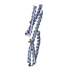

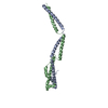
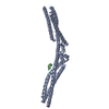


-モデル
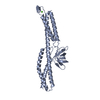
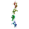
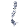
| モデル #218 |  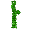 タイプ: dummy / ソフトウェア: DAMMIF / ダミー原子の半径: 3.00 A / カイ2乗値: 1.48328041  Omokage検索でこの集合体の類似形状データを探す (詳細) Omokage検索でこの集合体の類似形状データを探す (詳細) |
|---|
-試料
| 試料 | 名称: Plectin, fragment of the plakin domain encompassing the spectrin repeats SR3-SR4-SR5 and the SH3 試料濃度: 1.20-9.60 |
|---|---|
| バッファ | 名称: Sodium Phosphate / 濃度: 20.00 mM / pH: 7.5 / 組成: 150 mM NaCl, 5% glycerol, 2.5 mM DTT |
| 要素 #132 | タイプ: protein / 記述: Plectin / 分子量: 42.896 / 分子数: 1 / 由来: Homo sapiens / 参照: UniProt: Q15149 配列: ELEDSTLRYL QDLLAWVEEN QHRVDGAEWG VDLPSVEAQL GSHRGLHQSI EEFRAKIERA RSDEGQLSPA TRGAYRDCLG RLDLQYAKLL NSSKARLRSL ESLHSFVAAA TKELMWLNEK EEEEVGFDWS DRNTNMTAKK ESYSALMREL ELKEKKIKEL QNAGDRLLRE ...配列: ELEDSTLRYL QDLLAWVEEN QHRVDGAEWG VDLPSVEAQL GSHRGLHQSI EEFRAKIERA RSDEGQLSPA TRGAYRDCLG RLDLQYAKLL NSSKARLRSL ESLHSFVAAA TKELMWLNEK EEEEVGFDWS DRNTNMTAKK ESYSALMREL ELKEKKIKEL QNAGDRLLRE DHPARPTVES FQAALQTQWS WMLQLCCCIE AHLKENAAYF QFFSDVREAE GQLQKLQEAL RRKYSCDRSA TVTRLEDLLQ DAQDEKEQLN EYKGHLSGLA KRAKAVVQLK PRHPAHPMRG RLPLLAVCDY KQVEVTVHKG DECQLVGPAQ PSHWKVLSSS GSEAAVPSVC FLVPPPNQEA QEAVTRLEAQ HQALVTLWHQ LHVDMK |
-実験情報
| ビーム | 設備名称: Swiss Light Source cSAXS / 地域: Villigen / 国: Switzerland  / 線源: X-ray synchrotron / 波長: 0.1 Å / スペクトロメータ・検出器間距離: 2.15 mm / 線源: X-ray synchrotron / 波長: 0.1 Å / スペクトロメータ・検出器間距離: 2.15 mm | ||||||||||||||||||
|---|---|---|---|---|---|---|---|---|---|---|---|---|---|---|---|---|---|---|---|
| 検出器 | 名称: Pilatus 2M | ||||||||||||||||||
| スキャン | 
| ||||||||||||||||||
| 距離分布関数 P(R) | 
| ||||||||||||||||||
| 結果 |  コメント: Use of SAXS to analyze the structure of central region of the plakin domain of plectin.
|
