[English] 日本語
Yorodumi
- PDB-3pe0: Structure of the central region of the plakin domain of plectin -

+
Open data


ID or keywords:

Loading...

-
Basic information

Entry
Database: PDB / ID: 3pe0
TitleStructure of the central region of the plakin domain of plectin
ComponentsPlectin
KeywordsSTRUCTURAL PROTEIN / cytoskeleton / plakin / spectrin repeat / SH3 / intermediate filament / crosslinking
Function / homology
Function and homology information


protein-containing complex organization / actomyosin contractile ring assembly actin filament organization / skeletal myofibril assembly / tight junction organization / Type I hemidesmosome assembly / hemidesmosome assembly / hemidesmosome / leukocyte migration involved in immune response / intermediate filament organization / : ...protein-containing complex organization / actomyosin contractile ring assembly actin filament organization / skeletal myofibril assembly / tight junction organization / Type I hemidesmosome assembly / hemidesmosome assembly / hemidesmosome / leukocyte migration involved in immune response / intermediate filament organization / : / regulation of vascular permeability / intermediate filament cytoskeleton organization / dystroglycan binding / cellular response to hydrostatic pressure / fibroblast migration / costamere / T cell chemotaxis / cellular response to fluid shear stress / peripheral nervous system myelin maintenance / cardiac muscle cell development / myoblast differentiation / adherens junction organization / intermediate filament cytoskeleton / response to food / structural constituent of muscle / ankyrin binding / Assembly of collagen fibrils and other multimeric structures / intermediate filament / sarcomere organization / nucleus organization / keratinocyte development / transmission of nerve impulse / brush border / sarcoplasm / Caspase-mediated cleavage of cytoskeletal proteins / establishment of skin barrier / skeletal muscle fiber development / respiratory electron transport chain / mitochondrion organization / wound healing / cell morphogenesis / protein localization / multicellular organism growth / sarcolemma / structural constituent of cytoskeleton / Z disc / cellular response to mechanical stimulus / : / actin filament binding / myelin sheath / gene expression / mitochondrial outer membrane / cadherin binding / axon / focal adhesion / dendrite / perinuclear region of cytoplasm / RNA binding / extracellular exosome / plasma membrane / cytosol / cytoplasm
Similarity search - Function
: / Spectrin-like repeat / Spectrin repeat / Spectrin-like repeat / Methane Monooxygenase Hydroxylase; Chain G, domain 1 - #60 / Desmoplakin, spectrin-like domain / Spectrin like domain / Plectin repeat / Plectin repeat / Plakin repeat superfamily ...: / Spectrin-like repeat / Spectrin repeat / Spectrin-like repeat / Methane Monooxygenase Hydroxylase; Chain G, domain 1 - #60 / Desmoplakin, spectrin-like domain / Spectrin like domain / Plectin repeat / Plectin repeat / Plakin repeat superfamily / Desmoplakin, SH3 domain / SH3 domain / Plectin repeat / Plakin / Spectrin/alpha-actinin / Actinin-type actin-binding domain signature 1. / Actinin-type actin-binding domain signature 2. / Spectrin repeats / Actinin-type actin-binding domain, conserved site / Calponin homology domain / Calponin homology (CH) domain / Calponin homology domain / CH domain superfamily / Calponin homology (CH) domain profile. / SH3 Domains / Plectin/S10, N-terminal / Plectin/S10 domain / Methane Monooxygenase Hydroxylase; Chain G, domain 1 / SH3 type barrels. / Src homology 3 (SH3) domain profile. / SH3 domain / Roll / Winged helix-like DNA-binding domain superfamily / Up-down Bundle / Mainly Beta / Mainly Alpha
Similarity search - Domain/homology
Biological speciesHomo sapiens (human)
MethodX-RAY DIFFRACTION / SIRAS / Resolution: 2.95 Å
AuthorsOrtega, E. / de Pereda, J.M.
Citation
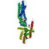
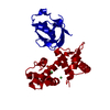
Journal: J Biol Chem / Year: 2011
Title: The structure of the plakin domain of plectin reveals a non-canonical SH3 domain interacting with its fourth spectrin repeat.
Authors: Esther Ortega / Rubén M Buey / Arnoud Sonnenberg / José M de Pereda /
Abstract: Plectin belongs to the plakin family of cytoskeletal crosslinkers, which is part of the spectrin superfamily. Plakins contain an N-terminal conserved region, the plakin domain, which is formed by an ...Plectin belongs to the plakin family of cytoskeletal crosslinkers, which is part of the spectrin superfamily. Plakins contain an N-terminal conserved region, the plakin domain, which is formed by an array of spectrin repeats (SR) and a Src-homology 3 (SH3), and harbors binding sites for junctional proteins. We have combined x-ray crystallography and small angle x-ray scattering (SAXS) to elucidate the structure of the central region of the plakin domain of plectin, which corresponds to the SR3, SR4, SR5, and SH3 domains. The crystal structures of the SR3-SR4 and SR4-SR5-SH3 fragments were determined to 2.2 and 2.95 Å resolution, respectively. The SH3 of plectin presents major alterations as compared with canonical Pro-rich binding SH3 domains, suggesting that plectin does not recognize Pro-rich motifs. In addition, the SH3 binding site is partially occluded by an intramolecular contact with the SR4. Residues of this pseudo-binding site and the SR4/SH3 interface are conserved within the plakin family, suggesting that the structure of this part of the plectin molecule is similar to that of other plakins. We have created a model for the SR3-SR4-SR5-SH3 region, which agrees well with SAXS data in solution. The three SRs form a semi-flexible rod that is not altered by the presence of the SH3 domain, and it is similar to those found in spectrins. The flexibility of the plakin domain, in analogy with spectrins, might contribute to the role of plakins in maintaining the stability of tissues subject to mechanical stress.
History
DepositionOct 25, 2010Deposition site: RCSB / Processing site: RCSB
Revision 1.0Feb 2, 2011Provider: repository / Type: Initial release
Revision 1.1Jul 13, 2011Group: Version format compliance
Revision 1.2Feb 21, 2024Group: Data collection / Database references / Derived calculations
Category: chem_comp_atom / chem_comp_bond ...chem_comp_atom / chem_comp_bond / database_2 / struct_conn / struct_ref_seq_dif / struct_site
Item: _database_2.pdbx_DOI / _database_2.pdbx_database_accession ..._database_2.pdbx_DOI / _database_2.pdbx_database_accession / _struct_conn.pdbx_dist_value / _struct_conn.ptnr1_auth_asym_id / _struct_conn.ptnr1_auth_comp_id / _struct_conn.ptnr1_auth_seq_id / _struct_conn.ptnr1_label_asym_id / _struct_conn.ptnr1_label_comp_id / _struct_conn.ptnr1_label_seq_id / _struct_conn.ptnr2_auth_asym_id / _struct_conn.ptnr2_label_asym_id / _struct_ref_seq_dif.details / _struct_site.pdbx_auth_asym_id / _struct_site.pdbx_auth_comp_id / _struct_site.pdbx_auth_seq_id

-
Structure visualization

Structure viewerMolecule:
MolmilJmol/JSmol

Downloads & links

-
Assembly

Deposited unit
A: Plectin
B: Plectin
hetero molecules


Theoretical massNumber of molelcules
Total (without water)64,7644
Polymers64,6832
Non-polymers802
Water0
1
A: Plectin
hetero molecules


Theoretical massNumber of molelcules
Total (without water)32,3822
Polymers32,3421
Non-polymers401
Water0
TypeNameSymmetry operationNumber
identity operation1_555x,y,z1
MethodPISA
2
B: Plectin
hetero molecules


Theoretical massNumber of molelcules
Total (without water)32,3822
Polymers32,3421
Non-polymers401
Water0
TypeNameSymmetry operationNumber
identity operation1_555x,y,z1
MethodPISA
Unit cell
Length a, b, c (Å)72.650, 108.512, 112.139
Angle α, β, γ (deg.)90.00, 90.00, 90.00
Int Tables number19
Space group name H-MP212121
Noncrystallographic symmetry (NCS)NCS domain:
IDEns-ID
11
21
12
22
13
23
14
24
15
25
16
26
17
27
18
28
19
29
110
210

NCS domain segments:
Dom-IDComponent-IDEns-IDSelection details
111chain B and resseq 642:668
211chain A and resseq 642:668
112chain B and resseq 669:678
212chain A and resseq 669:678
113chain A and resseq 679:713
213chain B and resseq 679:713
114chain B and resseq 714:745 and not (resid 724)
214chain A and resseq 714:745 and not (resid 724)
115chain B and (resseq 746:776 or resseq 795:824 or resseq 886:915) and not (resid 769)
215chain A and (resseq 746:776 or resseq 795:824 or resseq 886:915) and not (resid 769)
116chain A and resseq 777:782
216chain B and resseq 777:782
117chain B and (resseq 783:794 or resseq 1001)
217chain A and (resseq 783:794 or resseq 1001)
118chain A and (resseq 825:828)
218chain B and (resseq 825:828)
119chain A and (resseq 833:843 or resseq 848:870 or resseq 874:885) and not (resid 875)
219chain B and (resseq 833:843 or resseq 848:870 or resseq 874:885) and not (resid 875)
1110chain A and (resseq 844:847)
2110chain B and (resseq 844:847)

NCS ensembles :
ID
1
2
3
4
5
6
7
8
9
10

-
Components

#1: Protein Plectin / / PCN / PLTN / Hemidesmosomal protein 1 / HD1 / Plectin-1


Mass: 32341.748 Da / Num. of mol.: 2
Fragment: SPECTRIN REPEATS 4 AND 5, SH3 (UNP Residues 747-918)
Source method: isolated from a genetically manipulated source
Source: (gene. exp.) Homo sapiens (human) / Gene: PLEC, PLEC1 / Plasmid: Modified pET15b / Production host: Escherichia coli (E. coli) / Strain (production host): BL21(DE3) / References: UniProt: Q15149
#2: Chemical ChemComp-CA / CALCIUM ION


Mass: 40.078 Da / Num. of mol.: 2 / Source method: obtained synthetically / Formula: Ca

-
Experimental details

-
Experiment

ExperimentMethod: X-RAY DIFFRACTION / Number of used crystals: 1

-
Sample preparation

CrystalDensity Matthews: 3.42 Å3/Da / Density % sol: 64 %
Crystal growTemperature: 277 K / Method: vapor diffusion / pH: 7.2
Details: 0.1 M imidazole, 0.2 M calcium acetate, 9% PEG 8000, 2 mM DTT, pH 7.2, VAPOR DIFFUSION, temperature 277K

-
Data collection

DiffractionMean temperature: 100 K
Diffraction sourceSource: ROTATING ANODE / Type: BRUKER AXS MICROSTAR-H / Wavelength: 1.5418 Å
DetectorType: MAR scanner 345 mm plate / Detector: IMAGE PLATE / Date: Feb 12, 2010
RadiationMonochromator: HELIOS OPTICS / Protocol: SINGLE WAVELENGTH / Monochromatic (M) / Laue (L): M / Scattering type: x-ray
Radiation wavelengthWavelength: 1.5418 Å / Relative weight: 1
ReflectionResolution: 2.95→20 Å / Num. all: 18051 / Num. obs: 18051 / % possible obs: 93.6 % / Observed criterion σ(F): 0 / Observed criterion σ(I): 0 / Redundancy: 14.3 % / Biso Wilson estimate: 75.28 Å2 / Net I/σ(I): 29.4
Reflection shellResolution: 2.95→3.05 Å / Redundancy: 14.5 % / Mean I/σ(I) obs: 6.9 / % possible all: 85.5

-
Processing

Software
NameVersionClassification
MAR345dtbdata collection
SHELXmodel building
SHARPphasing
PHENIX(phenix.refine: 1.6.1_357)refinement
XDSdata reduction
XSCALEdata scaling
SHELXphasing
RefinementMethod to determine structure: SIRAS / Resolution: 2.95→19.977 Å / SU ML: 0.35 / σ(F): 1.8 / Phase error: 33.16 / Stereochemistry target values: ML
RfactorNum. reflection% reflectionSelection details
Rfree0.2654 925 5.13 %RANDOM
Rwork0.2413 ---
obs0.2426 18027 93.92 %-
all-18027 --
Solvent computationShrinkage radii: 0.9 Å / VDW probe radii: 1.11 Å / Solvent model: FLAT BULK SOLVENT MODEL / Bsol: 33.98 Å2 / ksol: 0.284 e/Å3
Displacement parameters
Baniso -1Baniso -2Baniso -3
1--8.4057 Å20 Å20 Å2
2--0.1991 Å20 Å2
3---8.2066 Å2
Refinement stepCycle: LAST / Resolution: 2.95→19.977 Å
ProteinNucleic acidLigandSolventTotal
Num. atoms4327 0 2 0 4329
Refine LS restraints
Refine-IDTypeDev idealNumber
X-RAY DIFFRACTIONf_bond_d0.0134870
X-RAY DIFFRACTIONf_angle_d1.0316289
X-RAY DIFFRACTIONf_dihedral_angle_d14.7331659
X-RAY DIFFRACTIONf_chiral_restr0.052656
X-RAY DIFFRACTIONf_plane_restr0.004780
Refine LS restraints NCS
Ens-IDDom-IDAuth asym-IDNumberRefine-IDTypeRms dev position (Å)
11B216X-RAY DIFFRACTIONPOSITIONAL
12A216X-RAY DIFFRACTIONPOSITIONAL0.033
21B89X-RAY DIFFRACTIONPOSITIONAL
22A89X-RAY DIFFRACTIONPOSITIONAL0.028
31A288X-RAY DIFFRACTIONPOSITIONAL
32B288X-RAY DIFFRACTIONPOSITIONAL0.03
41B248X-RAY DIFFRACTIONPOSITIONAL
42A248X-RAY DIFFRACTIONPOSITIONAL0.036
51B741X-RAY DIFFRACTIONPOSITIONAL
52A741X-RAY DIFFRACTIONPOSITIONAL0.046
61A42X-RAY DIFFRACTIONPOSITIONAL
62B42X-RAY DIFFRACTIONPOSITIONAL0.057
71B96X-RAY DIFFRACTIONPOSITIONAL
72A96X-RAY DIFFRACTIONPOSITIONAL0.044
81A32X-RAY DIFFRACTIONPOSITIONAL
82B32X-RAY DIFFRACTIONPOSITIONAL0.041
91A334X-RAY DIFFRACTIONPOSITIONAL
92B334X-RAY DIFFRACTIONPOSITIONAL0.041
101A28X-RAY DIFFRACTIONPOSITIONAL
102B28X-RAY DIFFRACTIONPOSITIONAL0.027
LS refinement shell
Resolution (Å)Rfactor RfreeNum. reflection RfreeRfactor RworkNum. reflection RworkRefine-ID% reflection obs (%)
2.95-3.1050.41281200.40712199X-RAY DIFFRACTION86
3.105-3.29880.41251300.34952387X-RAY DIFFRACTION93
3.2988-3.55210.34011470.30122440X-RAY DIFFRACTION96
3.5521-3.90720.30671270.25072491X-RAY DIFFRACTION96
3.9072-4.46710.25131360.21762409X-RAY DIFFRACTION93
4.4671-5.60730.17841210.19112502X-RAY DIFFRACTION95
5.6073-19.97730.20881440.20192674X-RAY DIFFRACTION98
Refinement TLS params.

Method: refined / Refine-ID: X-RAY DIFFRACTION

IDL112)L122)L132)L222)L232)L332)S11 (Å °)S12 (Å °)S13 (Å °)S21 (Å °)S22 (Å °)S23 (Å °)S31 (Å °)S32 (Å °)S33 (Å °)T112)T122)T132)T222)T232)T332)Origin x (Å)Origin y (Å)Origin z (Å)
11.8916-0.9993-1.60156.50863.3662.203-0.3664-0.54870.08680.28620.3940.0367-0.24250.4443-0.00230.35480.0295-0.08580.49290.1260.304576.916978.8806129.1797
20.0896-0.1551-0.18290.37190.49010.6038-0.25610.460.4516-0.9939-0.52840.3096-1.76370.1365-0.00120.9705-0.25410.08460.7430.02050.675580.704150.519391.7358
32.07990.2943-1.6367-0.0001-0.21121.18290.4396-0.47360.11620.28160.7196-0.75770.27330.1823-0.00170.57810.09610.00290.8796-0.22320.665890.327239.779980.8046
42.9915-1.17711.41184.20962.54323.7086-0.3840.0117-0.68150.2735-0.25841.06890.389-0.41580.00020.2853-0.13440.16230.2536-0.02510.755267.536758.8751119.2926
50.79550.44081.05370.37540.75011.5151-0.48330.1738-0.2922-0.0605-0.5440.6061-0.07841.3949-0.00010.711-0.0563-0.03140.6180.09180.771578.575839.699592.8276
62.2066-1.3814-2.33896.90584.25922.79750.0669-0.2516-0.0141-0.0574-0.07760.3873-0.2462-0.0380.00020.3524-0.0631-0.0730.25940.01280.166876.722278.804477.1722
70.1968-0.2736-0.24970.46730.54640.6084-0.2080.15670.3988-0.6448-0.62890.5186-1.7574-0.6898-0.00071.0293-0.06880.02680.7704-0.14550.513880.300550.497739.8233
81.9797-0.0805-1.568-0.02950.03791.20850.1587-0.58080.20670.1996-0.0218-0.01340.0419-0.4596-00.6357-0.10950.18210.7857-0.1270.557690.188939.818928.6771
93.7218-1.0220.41825.6971.73963.1261-0.22280.2188-0.47160.5148-1.26711.82640.3547-1.2372-0.00290.2886-0.22460.06310.3709-0.32990.656467.739358.413668.5228
100.80380.75780.55390.80320.85571.028-0.58050.4403-0.0782-0.2525-0.29960.8104-0.56160.95750.00070.571-0.17760.03440.6179-0.10840.569178.265939.918641.1863
Refinement TLS group
IDRefine-IDRefine TLS-IDSelection details
1X-RAY DIFFRACTION1(chain A and resid 642:753)
2X-RAY DIFFRACTION2(chain A and resid 754:774)
3X-RAY DIFFRACTION3(chain A and resid 775:804)
4X-RAY DIFFRACTION4(chain A and resid 805:892)
5X-RAY DIFFRACTION5(chain A and resid 893:915)
6X-RAY DIFFRACTION6(chain B and resid 642:753)
7X-RAY DIFFRACTION7(chain B and resid 754:774)
8X-RAY DIFFRACTION8(chain B and resid 775:804)
9X-RAY DIFFRACTION9(chain B and resid 805:891)
10X-RAY DIFFRACTION10(chain B and resid 892:915)

+
About Yorodumi

-
News

-
Feb 9, 2022. New format data for meta-information of EMDB entries

New format data for meta-information of EMDB entries

  • Version 3 of the EMDB header file is now the official format.
  • The previous official version 1.9 will be removed from the archive.

Related info.:EMDB header

External links:wwPDB to switch to version 3 of the EMDB data model

-
Aug 12, 2020. Covid-19 info

Covid-19 info

URL: https://pdbj.org/emnavi/covid19.php

New page: Covid-19 featured information page in EM Navigator.

Related info.:Covid-19 info / Mar 5, 2020. Novel coronavirus structure data

+
Mar 5, 2020. Novel coronavirus structure data

Novel coronavirus structure data

Related info.:Yorodumi Speices / Aug 12, 2020. Covid-19 info

External links:COVID-19 featured content - PDBj / Molecule of the Month (242):Coronavirus Proteases

+
Jan 31, 2019. EMDB accession codes are about to change! (news from PDBe EMDB page)

EMDB accession codes are about to change! (news from PDBe EMDB page)

  • The allocation of 4 digits for EMDB accession codes will soon come to an end. Whilst these codes will remain in use, new EMDB accession codes will include an additional digit and will expand incrementally as the available range of codes is exhausted. The current 4-digit format prefixed with “EMD-” (i.e. EMD-XXXX) will advance to a 5-digit format (i.e. EMD-XXXXX), and so on. It is currently estimated that the 4-digit codes will be depleted around Spring 2019, at which point the 5-digit format will come into force.
  • The EM Navigator/Yorodumi systems omit the EMD- prefix.

Related info.:Q: What is EMD? / ID/Accession-code notation in Yorodumi/EM Navigator

External links:EMDB Accession Codes are Changing Soon! / Contact to PDBj

+
Jul 12, 2017. Major update of PDB

Major update of PDB

  • wwPDB released updated PDB data conforming to the new PDBx/mmCIF dictionary.
  • This is a major update changing the version number from 4 to 5, and with Remediation, in which all the entries are updated.
  • In this update, many items about electron microscopy experimental information are reorganized (e.g. em_software).
  • Now, EM Navigator and Yorodumi are based on the updated data.

External links:wwPDB Remediation / Enriched Model Files Conforming to OneDep Data Standards Now Available in the PDB FTP Archive

-
Yorodumi

Thousand views of thousand structures

  • Yorodumi is a browser for structure data from EMDB, PDB, SASBDB, etc.
  • This page is also the successor to EM Navigator detail page, and also detail information page/front-end page for Omokage search.
  • The word "yorodu" (or yorozu) is an old Japanese word meaning "ten thousand". "mi" (miru) is to see.

Related info.:EMDB / PDB / SASBDB / Comparison of 3 databanks / Yorodumi Search / Aug 31, 2016. New EM Navigator & Yorodumi / Yorodumi Papers / Jmol/JSmol / Function and homology information / Changes in new EM Navigator and Yorodumi

Read more