 Movie
Movie Controller
[English] 日本語
 Yorodumi
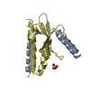
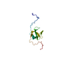
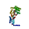
- SASDC84: parDE-like toxin-antitoxin module, EcPaaA2_13-63-HisEcParE2 construct -
+ Open data

- Basic information
| Entry | Database: SASBDB / ID: SASDC84 |
|---|---|
 Sample Sample | parDE-like toxin-antitoxin module, EcPaaA2_13-63-HisEcParE2 construct
|
| Function / homology | : / ParE toxin of type II toxin-antitoxin system, parDE / Toxin-antitoxin system, RelE/ParE toxin family / Toxin-antitoxin system, RelE/ParE toxin domain superfamily / Type II toxin-antitoxin system RelE/ParE family toxin / :  Function and homology information Function and homology information |
| Biological species |  |
 Citation Citation | Journal: J Mol Biol / Year: 2016 Title: A unique hetero-hexadecameric architecture displayed by the Escherichia coli O157 PaaA2-ParE2 antitoxin-toxin complex. Authors: Yann G-J Sterckx / Thomas Jové / Alexander V Shkumatov / Abel Garcia-Pino / Lieselotte Geerts / Maia De Kerpel / Jurij Lah / Henri De Greve / Laurence Van Melderen / Remy Loris /   Abstract: Many bacterial pathogens modulate their metabolic activity, virulence and pathogenicity through so-called "toxin-antitoxin" (TA) modules. The genome of the human pathogen Escherichia coli O157 ...Many bacterial pathogens modulate their metabolic activity, virulence and pathogenicity through so-called "toxin-antitoxin" (TA) modules. The genome of the human pathogen Escherichia coli O157 contains two three-component TA modules related to the known parDE module. Here, we show that the toxin EcParE2 maps in a branch of the RelE/ParE toxin superfamily that is distinct from the branches that contain verified gyrase and ribosome inhibitors. The structure of EcParE2 closely resembles that of Caulobacter crescentus ParE but shows a distinct pattern of conserved surface residues, in agreement with its apparent inability to interact with GyrA. The antitoxin EcPaaA2 is characterized by two α-helices (H1 and H2) that serve as molecular recognition elements to wrap itself around EcParE2. Both EcPaaA2 H1 and H2 are required to sustain a high-affinity interaction with EcParE2 and for the inhibition of EcParE2-mediated killing in vivo. Furthermore, evidence demonstrates that EcPaaA2 H2, but not H1, determines specificity for EcParE2. The initially formed EcPaaA2-EcParE2 heterodimer then assembles into a hetero-hexadecamer, which is stable in solution and is formed in a highly cooperative manner. Together these findings provide novel data on quaternary structure, TA interactions and activity of a hitherto poorly characterized family of TA modules. |
 Contact author Contact author |
|
- Structure visualization
| Structure viewer | Molecule: MolmilJmol/JSmol |
|---|
- Downloads & links
 SASDC84


-Models
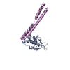
| Model #1318 |  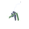 Type: atomic / Software: (r4556/NA) / Radius of dummy atoms: 1.90 A / Chi-square value: 0.760  Search similar-shape structures of this assembly by Omokage search (details) Search similar-shape structures of this assembly by Omokage search (details) |
|---|---|
| Model #1319 | 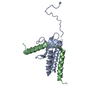 Type: atomic / Software: (r4556/NA) / Radius of dummy atoms: 1.90 A / Chi-square value: 0.760 Search similar-shape structures of this assembly by Omokage search (details) |
| Model #1320 |  Type: atomic / Software: (r8972/NA) / Radius of dummy atoms: 1.90 A / Chi-square value: 0.760 Search similar-shape structures of this assembly by Omokage search (details) |
-Sample
| Sample | Name: parDE-like toxin-antitoxin module, EcPaaA2_13-63-HisEcParE2 construct Specimen concentration: 12 mg/ml / Entity id: 702 / 704 |
|---|---|
| Buffer | Name: 50 mM Tris-HCl, 500 mM NaCl / pH: 7.5 |
| Entity #702 | Name: ParE2 / Type: protein / Description: Plasmid stabilization protein ParE / Formula weight: 12.596 / Num. of mol.: 1 / Source: Escherichia coli / References: UniProt: A0A0D7C2L1 Sequence: MGSSHHHHHH SSGLVPRGSH LPVLWLESAD TDLDDITSYI ARFDIDAAER LWQRLRGCVL PLSEHPYLYP PSDRVPGLRE IVAHPNYIIL YRVTTSSVEV VNVIHARRQF |
| Entity #704 | Name: PaaA2 / Type: protein / Description: Uncharacterized protein / Formula weight: 6.231 / Num. of mol.: 1 / Source: Escherichia coli O157:H7 str. SS52 / References: UniProt: A0A0F6F6Q9 Sequence: METIEQENSY NEWLRAKVAT SLADPRPAIP HDEVERRMAE RFAKMRKERS KQ |
-Experimental information
| Beam | Instrument name: SOLEIL SWING  / City: Saint-Aubin / 国: France / Type of source: X-ray synchrotron / Wavelength: 0.103 Å / Dist. spec. to detc.: 1.82 mm / City: Saint-Aubin / 国: France / Type of source: X-ray synchrotron / Wavelength: 0.103 Å / Dist. spec. to detc.: 1.82 mm | ||||||||||||||||||||||||||||||||||||
|---|---|---|---|---|---|---|---|---|---|---|---|---|---|---|---|---|---|---|---|---|---|---|---|---|---|---|---|---|---|---|---|---|---|---|---|---|---|
| Detector | Name: AVIEX / Type: CCD | ||||||||||||||||||||||||||||||||||||
| Scan |  Measurement date: Feb 5, 2012 / Storage temperature: 10 °C / Cell temperature: 10 °C / Exposure time: 0.75 sec. / Number of frames: 250 / Unit: 1/A /
| ||||||||||||||||||||||||||||||||||||
| Distance distribution function P(R) | 
| ||||||||||||||||||||||||||||||||||||
| Result | 
|
