 Movie
Movie Controller


+ Open data

- Basic information
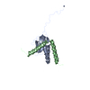
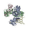
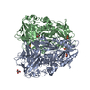
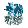
| Entry | Database: PDB / ID: 5cze | ||||||
|---|---|---|---|---|---|---|---|
| Title | Crystal structure of the PaaA2-ParE2 antitoxin-toxin complex | ||||||
 Components Components |
| ||||||
 Keywords Keywords | TOXIN / toxin-antitoxin | ||||||
| Function / homology |  Function and homology information Function and homology information: / ParE toxin of type II toxin-antitoxin system, parDE / RelE-like / Toxin-antitoxin system, RelE/ParE toxin family / YaeB-like fold / Toxin-antitoxin system, RelE/ParE toxin domain superfamily / 2-Layer Sandwich / Alpha Beta Similarity search - Domain/homology | ||||||
| Biological species |  | ||||||
| Method |  X-RAY DIFFRACTION / SYNCHROTRON / MOLECULAR REPLACEMENT / molecular replacement / Resolution: 3.82 Å X-RAY DIFFRACTION / SYNCHROTRON / MOLECULAR REPLACEMENT / molecular replacement / Resolution: 3.82 Å | ||||||
 Authors Authors | Loris, R. / Sterckx, Y.G.J. | ||||||
 Citation Citation | Journal: J Mol Biol / Year: 2016 Title: A unique hetero-hexadecameric architecture displayed by the Escherichia coli O157 PaaA2-ParE2 antitoxin-toxin complex. Authors: Yann G-J Sterckx / Thomas Jové / Alexander V Shkumatov / Abel Garcia-Pino / Lieselotte Geerts / Maia De Kerpel / Jurij Lah / Henri De Greve / Laurence Van Melderen / Remy Loris /   Abstract: Many bacterial pathogens modulate their metabolic activity, virulence and pathogenicity through so-called "toxin-antitoxin" (TA) modules. The genome of the human pathogen Escherichia coli O157 ...Many bacterial pathogens modulate their metabolic activity, virulence and pathogenicity through so-called "toxin-antitoxin" (TA) modules. The genome of the human pathogen Escherichia coli O157 contains two three-component TA modules related to the known parDE module. Here, we show that the toxin EcParE2 maps in a branch of the RelE/ParE toxin superfamily that is distinct from the branches that contain verified gyrase and ribosome inhibitors. The structure of EcParE2 closely resembles that of Caulobacter crescentus ParE but shows a distinct pattern of conserved surface residues, in agreement with its apparent inability to interact with GyrA. The antitoxin EcPaaA2 is characterized by two α-helices (H1 and H2) that serve as molecular recognition elements to wrap itself around EcParE2. Both EcPaaA2 H1 and H2 are required to sustain a high-affinity interaction with EcParE2 and for the inhibition of EcParE2-mediated killing in vivo. Furthermore, evidence demonstrates that EcPaaA2 H2, but not H1, determines specificity for EcParE2. The initially formed EcPaaA2-EcParE2 heterodimer then assembles into a hetero-hexadecamer, which is stable in solution and is formed in a highly cooperative manner. Together these findings provide novel data on quaternary structure, TA interactions and activity of a hitherto poorly characterized family of TA modules. | ||||||
| History |
|
- Structure visualization
| Structure viewer | Molecule: MolmilJmol/JSmol |
|---|
- Downloads & links
-Download
| PDBx/mmCIF format | 5cze.cif.gz | 266.8 KB | Display | PDBx/mmCIF format |
|---|---|---|---|---|
| PDB format | pdb5cze.ent.gz | 221 KB | Display | PDB format |
| PDBx/mmJSON format | 5cze.json.gz | Tree view | PDBx/mmJSON format | |
| Others |  Other downloads Other downloads |
-Validation report
| Arichive directory | https://data.pdbj.org/pub/pdb/validation_reports/cz/5czeftp://data.pdbj.org/pub/pdb/validation_reports/cz/5cze | HTTPS FTP |
|---|
-Related structure data
| Related structure data |  5cw7SC 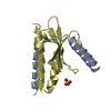 5czfC S: Starting model for refinement C: citing same article ( |
|---|---|
| Similar structure data |


-Links
 PDBj
PDBj- Assembly
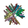
| Deposited unit | 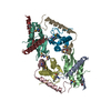
| ||||||||
|---|---|---|---|---|---|---|---|---|---|
| 1 | 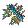
| ||||||||
| Unit cell |
|
-Components
| #1: Protein | Mass: 8661.044 Da / Num. of mol.: 4 Source method: isolated from a genetically manipulated source Source: (gene. exp.) Gene: SS52_2228 / Production host: #2: Protein | Mass: 11791.259 Da / Num. of mol.: 4 Source method: isolated from a genetically manipulated source Source: (gene. exp.) Gene: relE2, AML07_29260, APZ14_24120, ARC77_01680, ERS085383_05243, ERS085404_05085, ERS150873_04952, JEONG1266_09605 Production host: Has protein modification | Y | |
|---|
-Experimental details
-Experiment
| Experiment | Method: X-RAY DIFFRACTION / Number of used crystals: 1 |
|---|
- Sample preparation
| Crystal | Density Matthews: 3.17 Å3/Da / Density % sol: 61.22 % |
|---|---|
| Crystal grow | Temperature: 293 K / Method: vapor diffusion, hanging drop / pH: 4.6 Details: 20 mM calcium chloride, 100 mM sodium acetate pH 4.6, 30% MPD |
-Data collection
| Diffraction | Mean temperature: 100 K |
|---|---|
| Diffraction source | Source: SYNCHROTRON / Site: SOLEIL  / Beamline: PROXIMA 1 / Wavelength: 0.984 Å / Beamline: PROXIMA 1 / Wavelength: 0.984 Å |
| Detector | Type: DECTRIS PILATUS 6M / Detector: PIXEL / Date: Nov 24, 2010 |
| Radiation | Protocol: SINGLE WAVELENGTH / Monochromatic (M) / Laue (L): M / Scattering type: x-ray |
| Radiation wavelength | Wavelength: 0.984 Å / Relative weight: 1 |
| Reflection | Resolution: 3.822→46.88 Å / Num. obs: 9903 / % possible obs: 97.1 % / Redundancy: 7.8 % / Biso Wilson estimate: 124.93 Å2 / Rmerge(I) obs: 0.102 / Net I/σ(I): 12.91 |
| Reflection shell | Resolution: 3.82→3.96 Å / Redundancy: 6.6 % / Rmerge(I) obs: 0.494 / Mean I/σ(I) obs: 3.1 / Num. unique all: 841 / % possible all: 83.7 |
-Phasing
| Phasing | Method: molecular replacement |
|---|
- Processing
| Software |
| |||||||||||||||||||||||||||||||||||||||||||||||||||||||||||||||||||||||||||||||||||||||||||||||||||||||||||||||||||||||||||||||||||||||||||||||||||||||||||||||||||||||||||||||||||||||||||||||||||||||||||||||||||||||||||||||||
|---|---|---|---|---|---|---|---|---|---|---|---|---|---|---|---|---|---|---|---|---|---|---|---|---|---|---|---|---|---|---|---|---|---|---|---|---|---|---|---|---|---|---|---|---|---|---|---|---|---|---|---|---|---|---|---|---|---|---|---|---|---|---|---|---|---|---|---|---|---|---|---|---|---|---|---|---|---|---|---|---|---|---|---|---|---|---|---|---|---|---|---|---|---|---|---|---|---|---|---|---|---|---|---|---|---|---|---|---|---|---|---|---|---|---|---|---|---|---|---|---|---|---|---|---|---|---|---|---|---|---|---|---|---|---|---|---|---|---|---|---|---|---|---|---|---|---|---|---|---|---|---|---|---|---|---|---|---|---|---|---|---|---|---|---|---|---|---|---|---|---|---|---|---|---|---|---|---|---|---|---|---|---|---|---|---|---|---|---|---|---|---|---|---|---|---|---|---|---|---|---|---|---|---|---|---|---|---|---|---|---|---|---|---|---|---|---|---|---|---|---|---|---|---|---|---|---|
| Refinement | Method to determine structure: MOLECULAR REPLACEMENT Starting model: 5CW7 Resolution: 3.82→46.88 Å / Cor.coef. Fo:Fc: 0.805 / Cor.coef. Fo:Fc free: 0.796 / Rfactor Rfree error: 0 / Cross valid method: THROUGHOUT / σ(F): 0 / SU Rfree Blow DPI: 0.813
| |||||||||||||||||||||||||||||||||||||||||||||||||||||||||||||||||||||||||||||||||||||||||||||||||||||||||||||||||||||||||||||||||||||||||||||||||||||||||||||||||||||||||||||||||||||||||||||||||||||||||||||||||||||||||||||||||
| Displacement parameters | Biso mean: 113.38 Å2
| |||||||||||||||||||||||||||||||||||||||||||||||||||||||||||||||||||||||||||||||||||||||||||||||||||||||||||||||||||||||||||||||||||||||||||||||||||||||||||||||||||||||||||||||||||||||||||||||||||||||||||||||||||||||||||||||||
| Refine analyze | Luzzati coordinate error obs: 0.61 Å | |||||||||||||||||||||||||||||||||||||||||||||||||||||||||||||||||||||||||||||||||||||||||||||||||||||||||||||||||||||||||||||||||||||||||||||||||||||||||||||||||||||||||||||||||||||||||||||||||||||||||||||||||||||||||||||||||
| Refinement step | Cycle: LAST / Resolution: 3.82→46.88 Å
| |||||||||||||||||||||||||||||||||||||||||||||||||||||||||||||||||||||||||||||||||||||||||||||||||||||||||||||||||||||||||||||||||||||||||||||||||||||||||||||||||||||||||||||||||||||||||||||||||||||||||||||||||||||||||||||||||
| Refine LS restraints |
| |||||||||||||||||||||||||||||||||||||||||||||||||||||||||||||||||||||||||||||||||||||||||||||||||||||||||||||||||||||||||||||||||||||||||||||||||||||||||||||||||||||||||||||||||||||||||||||||||||||||||||||||||||||||||||||||||
| LS refinement shell | Resolution: 3.82→4.27 Å / Rfactor Rfree error: 0 / Total num. of bins used: 5
| |||||||||||||||||||||||||||||||||||||||||||||||||||||||||||||||||||||||||||||||||||||||||||||||||||||||||||||||||||||||||||||||||||||||||||||||||||||||||||||||||||||||||||||||||||||||||||||||||||||||||||||||||||||||||||||||||
| Refinement TLS params. | Method: refined / Refine-ID: X-RAY DIFFRACTION
| |||||||||||||||||||||||||||||||||||||||||||||||||||||||||||||||||||||||||||||||||||||||||||||||||||||||||||||||||||||||||||||||||||||||||||||||||||||||||||||||||||||||||||||||||||||||||||||||||||||||||||||||||||||||||||||||||
| Refinement TLS group |
|
