 Movie
Movie Controller

[English] 日本語
 Yorodumi
- PDB-3h25: Crystal structure of the catalytic domain of primase Repb' in com... -
+ Open data

- Basic information
| Entry | Database: PDB / ID: 3h25 | ||||||
|---|---|---|---|---|---|---|---|
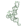
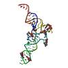
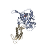
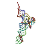
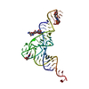
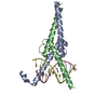
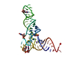
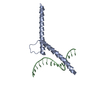
| Title | Crystal structure of the catalytic domain of primase Repb' in complex with initiator DNA | ||||||
 Components Components |
| ||||||
 Keywords Keywords | REPLICATION/DNA / PROTEIN-DNA-COMPLEX / HAIRPIN DNA / MIXED ALPHA-BETA FOLD / PRIMASE / RSF1010 / REPLICATION / REPLICATION-DNA COMPLEX | ||||||
| Function / homology |  Function and homology information Function and homology informationRepB-like DNA primase domain / : / RepB DNA-primase N-terminal domain / RepB DNA-primase C-terminal helical domain / Single helix bin / Single alpha-helices involved in coiled-coils or other helix-helix interfaces / Up-down Bundle / Mainly Alpha Similarity search - Domain/homology | ||||||
| Biological species | Plasmid RSF1010 (others) | ||||||
| Method |  X-RAY DIFFRACTION / SYNCHROTRON / MOLECULAR REPLACEMENT / Resolution: 2.7 Å X-RAY DIFFRACTION / SYNCHROTRON / MOLECULAR REPLACEMENT / Resolution: 2.7 Å | ||||||
 Authors Authors | Geibel, S. / Banchenko, S. / Engel, M. / Lanka, E. / Saenger, W. | ||||||
 Citation Citation | Journal: Proc.Natl.Acad.Sci.USA / Year: 2009 Title: Structure and function of primase RepB' encoded by broad-host-range plasmid RSF1010 that replicates exclusively in leading-strand mode Authors: Geibel, S. / Banchenko, S. / Engel, M. / Lanka, E. / Saenger, W. | ||||||
| History |
|
- Structure visualization
| Structure viewer | Molecule: MolmilJmol/JSmol |
|---|
- Downloads & links
-Download
| PDBx/mmCIF format | 3h25.cif.gz | 66.4 KB | Display | PDBx/mmCIF format |
|---|---|---|---|---|
| PDB format | pdb3h25.ent.gz | 44.8 KB | Display | PDB format |
| PDBx/mmJSON format | 3h25.json.gz | Tree view | PDBx/mmJSON format | |
| Others |  Other downloads Other downloads |
-Validation report
| Arichive directory | https://data.pdbj.org/pub/pdb/validation_reports/h2/3h25ftp://data.pdbj.org/pub/pdb/validation_reports/h2/3h25 | HTTPS FTP |
|---|
-Related structure data
| Related structure data | 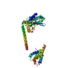 3h20SC S: Starting model for refinement C: citing same article ( |
|---|---|
| Similar structure data |
-Links
 PDBj
PDBj


- Assembly
| Deposited unit | 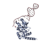
| ||||||||
|---|---|---|---|---|---|---|---|---|---|
| 1 |
| ||||||||
| Unit cell |
|
-Components
| #1: Protein | Mass: 25125.143 Da / Num. of mol.: 1 / Fragment: N-TERMINAL DOMAIN, UNP RESIDUES 1-212 Source method: isolated from a genetically manipulated source Source: (gene. exp.) Plasmid RSF1010 (others) / Gene: repb / Plasmid: pET28a / Production host:  |
|---|---|
| #2: DNA chain | Mass: 8270.298 Da / Num. of mol.: 1 / Fragment: SINGLE STRANDED INITIATOR DNA, GB 2701-2727 / Source method: obtained synthetically / Details: Synthetic DNA / References: GenBank: 152577 |
| #3: Water | ChemComp-HOH /  Mass: 18.015 Da / Num. of mol.: 24 / Source method: isolated from a natural source / Formula: H2O Mass: 18.015 Da / Num. of mol.: 24 / Source method: isolated from a natural source / Formula: H2O |
-Experimental details
-Experiment
| Experiment | Method: X-RAY DIFFRACTION / Number of used crystals: 1 |
|---|
- Sample preparation
| Crystal | Density Matthews: 1.88 Å3/Da / Density % sol: 34.52 % |
|---|---|
| Crystal grow | pH: 5 Details: 0.2M AMMONIUM CITRATE, 20% PEG 3350, VAPOR DIFFUSION, SITTING DROP, TEMPERATURE 291K |
-Data collection
| Diffraction | Mean temperature: 100 K |
|---|---|
| Diffraction source | Source: SYNCHROTRON / Site: BESSY  / Beamline: 14.2 / Wavelength: 0.91841 Å / Beamline: 14.2 / Wavelength: 0.91841 Å |
| Detector | Type: MAR CCD 165 mm / Detector: CCD / Date: Dec 5, 2007 / Details: MIRRORS |
| Radiation | Monochromator: DOUBLE CRYSTAL MONOCHROMATOR: SI-111 CRYSTAL / Protocol: SINGLE WAVELENGTH / Monochromatic (M) / Laue (L): M / Scattering type: x-ray |
| Radiation wavelength | Wavelength: 0.91841 Å / Relative weight: 1 |
| Reflection | Resolution: 2.7→19 Å / Num. all: 7400 / Num. obs: 7215 / % possible obs: 97.5 % / Observed criterion σ(I): 2 / Redundancy: 7.2 % / Rsym value: 0.111 / Net I/σ(I): 16.51 |
| Reflection shell | Resolution: 2.7→2.77 Å / Redundancy: 7.3 % / Mean I/σ(I) obs: 5.64 / Rsym value: 0.394 / % possible all: 95 |
- Processing
| Software |
| ||||||||||||||||||||||||||||||||||||||||||||||||||||||||||||||||||||||||||||||||||||||||||||||||||||||||||||||||||||||||||||||||||||||||||||||||||||||||||||||||||||||||||
|---|---|---|---|---|---|---|---|---|---|---|---|---|---|---|---|---|---|---|---|---|---|---|---|---|---|---|---|---|---|---|---|---|---|---|---|---|---|---|---|---|---|---|---|---|---|---|---|---|---|---|---|---|---|---|---|---|---|---|---|---|---|---|---|---|---|---|---|---|---|---|---|---|---|---|---|---|---|---|---|---|---|---|---|---|---|---|---|---|---|---|---|---|---|---|---|---|---|---|---|---|---|---|---|---|---|---|---|---|---|---|---|---|---|---|---|---|---|---|---|---|---|---|---|---|---|---|---|---|---|---|---|---|---|---|---|---|---|---|---|---|---|---|---|---|---|---|---|---|---|---|---|---|---|---|---|---|---|---|---|---|---|---|---|---|---|---|---|---|---|---|---|
| Refinement | Method to determine structure: MOLECULAR REPLACEMENT Starting model: CATALYTIC DOMAIN OF PDB 3H20, MODEL OF IDEAL B-DNA Resolution: 2.7→19 Å / Cor.coef. Fo:Fc: 0.908 / Cor.coef. Fo:Fc free: 0.863 / SU B: 30.309 / SU ML: 0.285 / TLS residual ADP flag: LIKELY RESIDUAL / Cross valid method: THROUGHOUT / ESU R Free: 0.401 / Stereochemistry target values: MAXIMUM LIKELIHOOD / Details: HYDROGENS HAVE BEEN ADDED IN THE RIDING POSITIONS
| ||||||||||||||||||||||||||||||||||||||||||||||||||||||||||||||||||||||||||||||||||||||||||||||||||||||||||||||||||||||||||||||||||||||||||||||||||||||||||||||||||||||||||
| Solvent computation | Ion probe radii: 0.8 Å / Shrinkage radii: 0.8 Å / VDW probe radii: 1.2 Å / Solvent model: BABINET MODEL WITH MASK | ||||||||||||||||||||||||||||||||||||||||||||||||||||||||||||||||||||||||||||||||||||||||||||||||||||||||||||||||||||||||||||||||||||||||||||||||||||||||||||||||||||||||||
| Displacement parameters | Biso mean: 31.47 Å2
| ||||||||||||||||||||||||||||||||||||||||||||||||||||||||||||||||||||||||||||||||||||||||||||||||||||||||||||||||||||||||||||||||||||||||||||||||||||||||||||||||||||||||||
| Refinement step | Cycle: LAST / Resolution: 2.7→19 Å
| ||||||||||||||||||||||||||||||||||||||||||||||||||||||||||||||||||||||||||||||||||||||||||||||||||||||||||||||||||||||||||||||||||||||||||||||||||||||||||||||||||||||||||
| Refine LS restraints |
| ||||||||||||||||||||||||||||||||||||||||||||||||||||||||||||||||||||||||||||||||||||||||||||||||||||||||||||||||||||||||||||||||||||||||||||||||||||||||||||||||||||||||||
| LS refinement shell | Resolution: 2.7→2.77 Å / Total num. of bins used: 20
| ||||||||||||||||||||||||||||||||||||||||||||||||||||||||||||||||||||||||||||||||||||||||||||||||||||||||||||||||||||||||||||||||||||||||||||||||||||||||||||||||||||||||||
| Refinement TLS params. | Method: refined / Origin x: 10.4478 Å / Origin y: 16.5103 Å / Origin z: -10.3449 Å
|
