 Movie
Movie Controller

+ Open data

- Basic information
| Entry | Database: SASBDB / ID: SASDC54 |
|---|---|
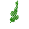
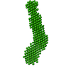
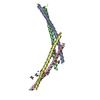
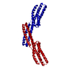
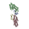
 Sample Sample | Envelope of Col H PKD-PKD-CBD complexed with mini-collagen
|
| Function / homology |  Function and homology information Function and homology informationtripeptidase activity / microbial collagenase / collagen metabolic process / collagen binding / metalloendopeptidase activity / endopeptidase activity / calcium ion binding / proteolysis / extracellular region / zinc ion binding Similarity search - Function |
| Biological species | Hathewaya histolytica (Clostridium histolyticum) synthetic construct (others) |
 Citation Citation | Journal: FEBS J / Year: 2018 Title: Ca -induced orientation of tandem collagen binding domains from clostridial collagenase ColG permits two opposing functions of collagen fibril formation and retardation. Authors: Perry Caviness / Ryan Bauer / Keisuke Tanaka / Katarzyna Janowska / Jeffrey Randall Roeser / Dawn Harter / Jes Sanders / Christopher Ruth / Osamu Matsushita / Joshua Sakon /   Abstract: To penetrate host tissues, histotoxic clostridia secrete virulence factors including enzymes to hydrolyze extracellular matrix. Clostridium histolyticum, recently renamed as Hathewaya histolytica, ...To penetrate host tissues, histotoxic clostridia secrete virulence factors including enzymes to hydrolyze extracellular matrix. Clostridium histolyticum, recently renamed as Hathewaya histolytica, produces two classes of collagenase (ColG and ColH). The high-speed AFM study showed that ColG collagenase moves unidirectionally to plane collagen fibril and rebundles fibril when stalled . The structural explanation of the roles for the tandem collagen-binding segment (CBDs) is illuminated by its calcium-bound crystal structure at 1.9 Å resolution (R = 15.0%; R = 19.6%). Activation may involve calcium-dependent domain rearrangement supported by both small-angle X-ray scattering and size exclusion chromatography. At pCa ≥ 5 (pCa = -log[Ca ]), the tandem CBD adopts an extended conformation that may facilitate secretion from the bacterium. At pCa ≤ 4, the compact structure seen in the crystal structure is adopted. This arrangement positions the two binding surfaces ~ 55 Å apart, and possibly ushers ColG along tropocollagen molecules that allow for unidirectional movement. A sequential binding mode where tighter binding CBD2 binds first could aid in processivity as well. Switch from processive collagenolysis to fibril rearrangement could be concentration dependent. Collagen fibril formation is retarded at 1 : 1 molar ratio of tandem CBD to collagen. Tandem CBD may help isolate a tropocollagen molecule from a fibril at this ratio. At 0.1 : 1 to 0.5 : 1 molar ratios fibril self-assembly was accelerated. Gain of function as a result of gene duplication of CBD for the M9B enzymes is speculated. The binding and activation modes described here will aid in drug delivery design. ACCESSION CODES: The full atomic coordinates of the tandem CBD and its corresponding structure factor amplitudes have been deposited in the Protein Data Bank (PDB accession code 5IKU). Small-angle X- ...ACCESSION CODES: The full atomic coordinates of the tandem CBD and its corresponding structure factor amplitudes have been deposited in the Protein Data Bank (PDB accession code 5IKU). Small-angle X-ray scattering data and corresponding ab initio models have been submitted to the Small Angle Scattering Biological Data Bank (SASBDB). Accession codes CL2, collagenase module 2, CN2, CP2 are assigned to envelopes for tandem CBD at -log[Ca ] (pCa) 3, 4, 5, and 6, respectively. Accession code DC64 was assigned to the complex of polycystic kidney disease-CBD1-CBD2 with mini-collagen. |
 Contact author Contact author |
|
- Structure visualization
| Structure viewer | Molecule: MolmilJmol/JSmol |
|---|
- Downloads & links
 SASDC54
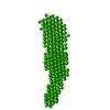


-Models
| Model #1285 |   Type: dummy / Radius of dummy atoms: 2.50 A / Chi-square value: 3.217 / P-value: 0.000003  Search similar-shape structures of this assembly by Omokage search (details) Search similar-shape structures of this assembly by Omokage search (details) |
|---|
-Sample
| Sample | Name: Envelope of Col H PKD-PKD-CBD complexed with mini-collagen Specimen concentration: 1.00-5.00 / Entity id: 675 / 677 |
|---|---|
| Buffer | Name: 50 mM Hepes 100 mM NaCl 5 mM CaCl2 / pH: 7.5 |
| Entity #675 | Name: ColH / Type: protein / Description: ColH protein / Formula weight: 33.727 / Num. of mol.: 1 / Source: Hathewaya histolytica (Clostridium histolyticum) / References: UniProt: Q46085 Sequence: lpnegdskns lpygkingty kgtekekikf ssegsfdpdg kivsyewdfg dgnksneenp ehsydkvgty tvklkvtddk gessvsttta eikdlsenkl pviymhvpks galnqkvvfy gkgtydpdgs iagyqwdfgd gsdfsseqnp shvytkkgey tvtlrvmdss ...Sequence: lpnegdskns lpygkingty kgtekekikf ssegsfdpdg kivsyewdfg dgnksneenp ehsydkvgty tvklkvtddk gessvsttta eikdlsenkl pviymhvpks galnqkvvfy gkgtydpdgs iagyqwdfgd gsdfsseqnp shvytkkgey tvtlrvmdss gqmsektmki kitdpvypig tekepnnske tasgpivpgi pvsgtients dqdyfyfdvi tpgevkidin klgyggatwv vydennnavs yatddgqnls gkfkadkpgr yyihlymfng sympyrinie gsvgr |
| Entity #677 | Type: protein / Description: Collagenous Peptide model [(PPG)10] / Formula weight: 3.21 / Num. of mol.: 3 / Source: synthetic construct Sequence: PPGPPGPPGP PGPPGPPGPP GPPGPPGPPG |
-Experimental information
| Beam | Instrument name: Advanced Light Source (ALS) 12.3.1 (SIBYLS) City: Berkeley, CA / 国: USA / Type of source: X-ray synchrotron / Wavelength: 0.113 Å | |||||||||||||||||||||||||||||||||
|---|---|---|---|---|---|---|---|---|---|---|---|---|---|---|---|---|---|---|---|---|---|---|---|---|---|---|---|---|---|---|---|---|---|---|
| Detector | Name: Pilatus3 X 2M / Pixsize x: 172 mm | |||||||||||||||||||||||||||||||||
| Scan |  Measurement date: Oct 12, 2016 / Storage temperature: 4 °C / Cell temperature: 10 °C / Exposure time: 0.3 sec. / Number of frames: 33 / Unit: 1/A /
| |||||||||||||||||||||||||||||||||
| Distance distribution function P(R) | 
| |||||||||||||||||||||||||||||||||
| Result |  Comments: Scattering for samples with concentrations of 4 mg/ml, 3 mg/ml and 1 mg/ml were averaged. Each of the average scattering curves are used to extrapolate towards infinite dilution. The ...Comments: Scattering for samples with concentrations of 4 mg/ml, 3 mg/ml and 1 mg/ml were averaged. Each of the average scattering curves are used to extrapolate towards infinite dilution. The scattering curve at infinite dilution was used to generate the SAXS model that represents the averaged spatial disposition of the complex in solution (DAMFILT).
|
