[English] 日本語
Yorodumi
- PDB-5iku: Crystal structure of the Hathewaya histolytica ColG tandem collag... -

+
Open data


ID or keywords:

Loading...

-
Basic information

Entry
Database: PDB / ID: 5iku
TitleCrystal structure of the Hathewaya histolytica ColG tandem collagen-binding domain s3as3b in the presence of calcium at 1.9 Angstrom resolution
ComponentsCollagenase
KeywordsPROTEIN BINDING / Calcium-binding protein Collagen-binding protein
Function / homology
Function and homology information


tripeptidase activity / microbial collagenase / collagen metabolic process / collagen binding / metalloendopeptidase activity / endopeptidase activity / calcium ion binding / proteolysis / zinc ion binding / extracellular region
Similarity search - Function
Collagenase ColG, catalytic helper subdomain / Collagenase G catalytic helper subdomain / Peptidase M9A/M9B, collagenase, bacterial / Peptidase M9, collagenase, N-terminal domain / Collagenase / Peptidase family M9 N-terminal / Peptidase, C-terminal, archaeal/bacterial / Bacterial pre-peptidase C-terminal domain / PKD domain / Polycystic kidney disease (PKD) domain profile. ...Collagenase ColG, catalytic helper subdomain / Collagenase G catalytic helper subdomain / Peptidase M9A/M9B, collagenase, bacterial / Peptidase M9, collagenase, N-terminal domain / Collagenase / Peptidase family M9 N-terminal / Peptidase, C-terminal, archaeal/bacterial / Bacterial pre-peptidase C-terminal domain / PKD domain / Polycystic kidney disease (PKD) domain profile. / PKD domain / PKD domain superfamily / PKD/Chitinase domain / Repeats in polycystic kidney disease 1 (PKD1) and other proteins / Neutral zinc metallopeptidases, zinc-binding region signature. / Immunoglobulin-like fold
Similarity search - Domain/homology
Biological speciesHathewaya histolytica (bacteria)
MethodX-RAY DIFFRACTION / Resolution: 1.9 Å
AuthorsJanowska, K. / Bauer, R. / Roeser, R. / Sakon, J. / Matsushita, O.
Funding support United States, 1items
OrganizationGrant numberCountry
National Institutes of Health/National Institute of General Medical Sciences (NIH/NIGMS)8P30GM103450 United States
Citation
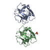
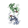
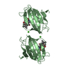
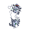
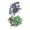
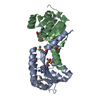
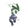
Journal: FEBS J / Year: 2018
Title: Ca -induced orientation of tandem collagen binding domains from clostridial collagenase ColG permits two opposing functions of collagen fibril formation and retardation.
Authors: Perry Caviness / Ryan Bauer / Keisuke Tanaka / Katarzyna Janowska / Jeffrey Randall Roeser / Dawn Harter / Jes Sanders / Christopher Ruth / Osamu Matsushita / Joshua Sakon /
Abstract: To penetrate host tissues, histotoxic clostridia secrete virulence factors including enzymes to hydrolyze extracellular matrix. Clostridium histolyticum, recently renamed as Hathewaya histolytica, ...To penetrate host tissues, histotoxic clostridia secrete virulence factors including enzymes to hydrolyze extracellular matrix. Clostridium histolyticum, recently renamed as Hathewaya histolytica, produces two classes of collagenase (ColG and ColH). The high-speed AFM study showed that ColG collagenase moves unidirectionally to plane collagen fibril and rebundles fibril when stalled . The structural explanation of the roles for the tandem collagen-binding segment (CBDs) is illuminated by its calcium-bound crystal structure at 1.9 Å resolution (R = 15.0%; R = 19.6%). Activation may involve calcium-dependent domain rearrangement supported by both small-angle X-ray scattering and size exclusion chromatography. At pCa ≥ 5 (pCa = -log[Ca ]), the tandem CBD adopts an extended conformation that may facilitate secretion from the bacterium. At pCa ≤ 4, the compact structure seen in the crystal structure is adopted. This arrangement positions the two binding surfaces ~ 55 Å apart, and possibly ushers ColG along tropocollagen molecules that allow for unidirectional movement. A sequential binding mode where tighter binding CBD2 binds first could aid in processivity as well. Switch from processive collagenolysis to fibril rearrangement could be concentration dependent. Collagen fibril formation is retarded at 1 : 1 molar ratio of tandem CBD to collagen. Tandem CBD may help isolate a tropocollagen molecule from a fibril at this ratio. At 0.1 : 1 to 0.5 : 1 molar ratios fibril self-assembly was accelerated. Gain of function as a result of gene duplication of CBD for the M9B enzymes is speculated. The binding and activation modes described here will aid in drug delivery design.
ACCESSION CODES: The full atomic coordinates of the tandem CBD and its corresponding structure factor amplitudes have been deposited in the Protein Data Bank (PDB accession code 5IKU). Small-angle X- ...ACCESSION CODES: The full atomic coordinates of the tandem CBD and its corresponding structure factor amplitudes have been deposited in the Protein Data Bank (PDB accession code 5IKU). Small-angle X-ray scattering data and corresponding ab initio models have been submitted to the Small Angle Scattering Biological Data Bank (SASBDB). Accession codes CL2, collagenase module 2, CN2, CP2 are assigned to envelopes for tandem CBD at -log[Ca ] (pCa) 3, 4, 5, and 6, respectively. Accession code DC64 was assigned to the complex of polycystic kidney disease-CBD1-CBD2 with mini-collagen.
History
DepositionMar 3, 2016Deposition site: RCSB / Processing site: RCSB
Revision 1.0Mar 15, 2017Provider: repository / Type: Initial release
Revision 1.1Sep 20, 2017Group: Author supporting evidence / Refinement description / Category: pdbx_audit_support / software / Item: _pdbx_audit_support.funding_organization
Revision 1.2Dec 25, 2019Group: Author supporting evidence / Category: pdbx_audit_support / Item: _pdbx_audit_support.funding_organization
Revision 1.3Feb 19, 2020Group: Database references / Source and taxonomy / Structure summary
Category: citation / citation_author ...citation / citation_author / entity_src_gen / struct
Item: _citation.country / _citation.journal_abbrev ..._citation.country / _citation.journal_abbrev / _citation.journal_id_CSD / _citation.journal_id_ISSN / _citation.journal_volume / _citation.page_first / _citation.page_last / _citation.pdbx_database_id_DOI / _citation.pdbx_database_id_PubMed / _citation.title / _citation.year / _entity_src_gen.pdbx_gene_src_scientific_name / _struct.title
Revision 1.4Mar 6, 2024Group: Data collection / Database references / Category: chem_comp_atom / chem_comp_bond / database_2
Item: _database_2.pdbx_DOI / _database_2.pdbx_database_accession

-
Structure visualization

Structure viewerMolecule:
MolmilJmol/JSmol

Downloads & links

-
Assembly

Deposited unit
A: Collagenase
hetero molecules


Theoretical massNumber of molelcules
Total (without water)27,0085
Polymers26,8471
Non-polymers1604
Water3,801211
1


  • Idetical with deposited unit
  • defined by author&software
TypeNameSymmetry operationNumber
identity operation1_555x,y,z1
MethodPISA
Unit cell
Length a, b, c (Å)51.520, 54.710, 92.040
Angle α, β, γ (deg.)90.000, 90.000, 90.000
Int Tables number19
Space group name H-MP212121

-
Components

#1: Protein Collagenase


Mass: 26847.432 Da / Num. of mol.: 1 / Fragment: unp residues 883-1118
Source method: isolated from a genetically manipulated source
Source: (gene. exp.) Hathewaya histolytica (bacteria) / Gene: colG / Production host: Escherichia coli (E. coli) / Strain (production host): BL21 (RIL) / References: UniProt: Q9X721
#2: Chemical
ChemComp-CA / CALCIUM ION


Mass: 40.078 Da / Num. of mol.: 4 / Source method: obtained synthetically / Formula: Ca
#3: Water ChemComp-HOH / water


Mass: 18.015 Da / Num. of mol.: 211 / Source method: isolated from a natural source / Formula: H2O

-
Experimental details

-
Experiment

ExperimentMethod: X-RAY DIFFRACTION / Number of used crystals: 1

-
Sample preparation

CrystalDensity Matthews: 2.42 Å3/Da / Density % sol: 49.08 %
Crystal growTemperature: 310 K / Method: vapor diffusion, hanging drop / pH: 7.5
Details: 0.003 M calcium chloride, 0.1 M Bis-Tris pH 7.5, and 21-26% (w/v) PEG 3350.

-
Data collection

DiffractionMean temperature: 296 K
Diffraction sourceSource: ROTATING ANODE / Type: RIGAKU MICROMAX-007 / Wavelength: 1.5418 Å
DetectorType: RIGAKU / Detector: IMAGE PLATE / Date: Feb 6, 2012 / Details: CCD
RadiationMonochromator: Osmic Blue confocal mirrors / Protocol: SINGLE WAVELENGTH / Scattering type: x-ray
Radiation wavelengthWavelength: 1.5418 Å / Relative weight: 1
ReflectionResolution: 1.9→47.03 Å / Num. obs: 20021 / % possible obs: 94.8 % / Redundancy: 4.47 % / Biso Wilson estimate: 37.7 Å2 / Rmerge(I) obs: 0.063 / Net I/σ(I): 11.2
Reflection shellResolution: 1.9→1.97 Å / Redundancy: 4.27 % / Rmerge(I) obs: 0.425 / Mean I/σ(I) obs: 2.9 / % possible all: 94.2

-
Processing

Software
NameVersionClassification
d*TREK9.9.9.4 W9RSSIdata scaling
REFMAC5.8.0135refinement
PDB_EXTRACT3.2data extraction
d*TREKdata reduction
MOLREPphasing
RefinementResolution: 1.9→47.03 Å / Cor.coef. Fo:Fc: 0.981 / Cor.coef. Fo:Fc free: 0.968 / SU B: 6.547 / SU ML: 0.099 / Cross valid method: THROUGHOUT / σ(F): 0 / ESU R: 0.133 / ESU R Free: 0.129 / Details: U VALUES : WITH TLS ADDED
RfactorNum. reflection% reflectionSelection details
Rfree0.1963 1027 5.1 %RANDOM
Rwork0.1497 ---
obs0.152 18987 94.65 %-
Solvent computationIon probe radii: 0.8 Å / Shrinkage radii: 0.8 Å / VDW probe radii: 1.2 Å
Displacement parametersBiso max: 181.64 Å2 / Biso mean: 47.397 Å2 / Biso min: 21.44 Å2
Baniso -1Baniso -2Baniso -3
1-1.05 Å20 Å2-0 Å2
2---0.8 Å20 Å2
3----0.24 Å2
Refinement stepCycle: final / Resolution: 1.9→47.03 Å
ProteinNucleic acidLigandSolventTotal
Num. atoms1892 0 4 211 2107
Biso mean--37.26 64.69 -
Num. residues----240
Refine LS restraints
Refine-IDTypeDev idealDev ideal targetNumber
X-RAY DIFFRACTIONr_bond_refined_d0.0110.0191951
X-RAY DIFFRACTIONr_angle_refined_deg1.8661.9522632
X-RAY DIFFRACTIONr_dihedral_angle_1_deg6.1255239
X-RAY DIFFRACTIONr_dihedral_angle_2_deg35.64725.9100
X-RAY DIFFRACTIONr_dihedral_angle_3_deg14.63415348
X-RAY DIFFRACTIONr_dihedral_angle_4_deg14.213156
X-RAY DIFFRACTIONr_chiral_restr0.1660.2279
X-RAY DIFFRACTIONr_gen_planes_refined0.0060.021490
X-RAY DIFFRACTIONr_mcbond_it3.8053.089959
X-RAY DIFFRACTIONr_mcangle_it4.7194.5961197
X-RAY DIFFRACTIONr_scbond_it6.8353.67992
LS refinement shellResolution: 1.9→1.949 Å / Total num. of bins used: 20
RfactorNum. reflection% reflection
Rfree0.268 66 -
Rwork0.255 1382 -
all-1448 -
obs--93.72 %
Refinement TLS params.

Method: refined / Refine-ID: X-RAY DIFFRACTION

IDL112)L122)L132)L222)L232)L332)S11 (Å °)S12 (Å °)S13 (Å °)S21 (Å °)S22 (Å °)S23 (Å °)S31 (Å °)S32 (Å °)S33 (Å °)T112)T122)T132)T222)T232)T332)Origin x (Å)Origin y (Å)Origin z (Å)
11.69970.2849-0.63353.2193-0.32481.54080.00460.12080.1224-0.2236-0.0004-0.1124-0.01210.0224-0.00420.04040.01080.02120.02420.02430.0294-21.576322.1395-28.7982
23.09650.28540.78033.4020.51742.85750.05340.0313-0.10810.11710.0814-0.19330.03740.2439-0.13480.10340.0067-0.07140.0580.00130.0635-8.72353.3902-2.5693
Refinement TLS group
IDRefine-IDRefine TLS-IDAuth asym-IDAuth seq-ID
1X-RAY DIFFRACTION1A778 - 890
2X-RAY DIFFRACTION2A897 - 1007

+
About Yorodumi

-
News

-
Feb 9, 2022. New format data for meta-information of EMDB entries

New format data for meta-information of EMDB entries

  • Version 3 of the EMDB header file is now the official format.
  • The previous official version 1.9 will be removed from the archive.

Related info.:EMDB header

External links:wwPDB to switch to version 3 of the EMDB data model

-
Aug 12, 2020. Covid-19 info

Covid-19 info

URL: https://pdbjlc1.pdbj.org/emnavi/covid19.php

New page: Covid-19 featured information page in EM Navigator.

Related info.:Covid-19 info / Mar 5, 2020. Novel coronavirus structure data

+
Mar 5, 2020. Novel coronavirus structure data

Novel coronavirus structure data

Related info.:Yorodumi Speices / Aug 12, 2020. Covid-19 info

External links:COVID-19 featured content - PDBj / Molecule of the Month (242):Coronavirus Proteases

+
Jan 31, 2019. EMDB accession codes are about to change! (news from PDBe EMDB page)

EMDB accession codes are about to change! (news from PDBe EMDB page)

  • The allocation of 4 digits for EMDB accession codes will soon come to an end. Whilst these codes will remain in use, new EMDB accession codes will include an additional digit and will expand incrementally as the available range of codes is exhausted. The current 4-digit format prefixed with “EMD-” (i.e. EMD-XXXX) will advance to a 5-digit format (i.e. EMD-XXXXX), and so on. It is currently estimated that the 4-digit codes will be depleted around Spring 2019, at which point the 5-digit format will come into force.
  • The EM Navigator/Yorodumi systems omit the EMD- prefix.

Related info.:Q: What is EMD? / ID/Accession-code notation in Yorodumi/EM Navigator

External links:EMDB Accession Codes are Changing Soon! / Contact to PDBj

+
Jul 12, 2017. Major update of PDB

Major update of PDB

  • wwPDB released updated PDB data conforming to the new PDBx/mmCIF dictionary.
  • This is a major update changing the version number from 4 to 5, and with Remediation, in which all the entries are updated.
  • In this update, many items about electron microscopy experimental information are reorganized (e.g. em_software).
  • Now, EM Navigator and Yorodumi are based on the updated data.

External links:wwPDB Remediation / Enriched Model Files Conforming to OneDep Data Standards Now Available in the PDB FTP Archive

-
Yorodumi

Thousand views of thousand structures

  • Yorodumi is a browser for structure data from EMDB, PDB, SASBDB, etc.
  • This page is also the successor to EM Navigator detail page, and also detail information page/front-end page for Omokage search.
  • The word "yorodu" (or yorozu) is an old Japanese word meaning "ten thousand". "mi" (miru) is to see.

Related info.:EMDB / PDB / SASBDB / Comparison of 3 databanks / Yorodumi Search / Aug 31, 2016. New EM Navigator & Yorodumi / Yorodumi Papers / Jmol/JSmol / Function and homology information / Changes in new EM Navigator and Yorodumi

Read more