 Movie
Movie Controller

[English] 日本語
 Yorodumi
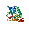
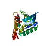
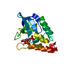
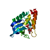
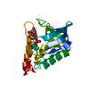
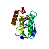
- PDB-3em0: Crystal structure of Zebrafish Ileal Bile Acid-Bindin Protein com... -
+ Open data

- Basic information
| Entry | Database: PDB / ID: 3em0 | ||||||
|---|---|---|---|---|---|---|---|
| Title | Crystal structure of Zebrafish Ileal Bile Acid-Bindin Protein complexed with cholic acid (crystal form B). | ||||||
 Components Components | Ileal Bile Acid-Binding Protein | ||||||
 Keywords Keywords | LIPID BINDING PROTEIN / Ileal bile acid-binding protein / zebrafish (Danio rerio) / cholic acid / Lipid-binding / Transport | ||||||
| Function / homology |  Function and homology information Function and homology informationRecycling of bile acids and salts / Triglyceride catabolism / bile acid binding / fatty acid transport / fatty acid binding / nucleus / cytosol Similarity search - Function | ||||||
| Biological species |  | ||||||
| Method |  X-RAY DIFFRACTION / SYNCHROTRON / MOLECULAR REPLACEMENT / Resolution: 2.2 Å X-RAY DIFFRACTION / SYNCHROTRON / MOLECULAR REPLACEMENT / Resolution: 2.2 Å | ||||||
 Authors Authors | Capaldi, S. / Saccomani, G. / Fessas, D. / Signorelli, M. / Perduca, M. / Monaco, H.L. | ||||||
 Citation Citation | Journal: J.Mol.Biol. / Year: 2009 Title: The X-Ray structure of zebrafish (Danio rerio) ileal bile acid-binding protein reveals the presence of binding sites on the surface of the protein molecule. Authors: Capaldi, S. / Saccomani, G. / Fessas, D. / Signorelli, M. / Perduca, M. / Monaco, H.L. | ||||||
| History |
|
- Structure visualization
| Structure viewer | Molecule: MolmilJmol/JSmol |
|---|
- Downloads & links
-Download
| PDBx/mmCIF format | 3em0.cif.gz | 71.8 KB | Display | PDBx/mmCIF format |
|---|---|---|---|---|
| PDB format | pdb3em0.ent.gz | 54.2 KB | Display | PDB format |
| PDBx/mmJSON format | 3em0.json.gz | Tree view | PDBx/mmJSON format | |
| Others |  Other downloads Other downloads |
-Validation report
| Arichive directory | https://data.pdbj.org/pub/pdb/validation_reports/em/3em0ftp://data.pdbj.org/pub/pdb/validation_reports/em/3em0 | HTTPS FTP |
|---|
-Related structure data
| Related structure data | 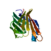 3elxC 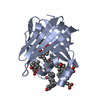 3elzSC C: citing same article ( S: Starting model for refinement |
|---|---|
| Similar structure data |

-Links
 PDBj
PDBj

- Assembly
| Deposited unit | 
| ||||||||
|---|---|---|---|---|---|---|---|---|---|
| 1 | 
| ||||||||
| 2 | 
| ||||||||
| Unit cell |
| ||||||||
| Details | The biological assembly is the protein monomer. |
-Components
| #1: Protein | Mass: 15191.230 Da / Num. of mol.: 2 Source method: isolated from a genetically manipulated source Source: (gene. exp.)  #2: Chemical | ChemComp-CHD / 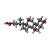  Mass: 408.571 Da / Num. of mol.: 9 / Source method: obtained synthetically / Formula: C24H40O5 Mass: 408.571 Da / Num. of mol.: 9 / Source method: obtained synthetically / Formula: C24H40O5#3: Water | ChemComp-HOH / |  Mass: 18.015 Da / Num. of mol.: 69 / Source method: isolated from a natural source / Formula: H2O Mass: 18.015 Da / Num. of mol.: 69 / Source method: isolated from a natural source / Formula: H2O |
|---|
-Experimental details
-Experiment
| Experiment | Method: X-RAY DIFFRACTION / Number of used crystals: 1 |
|---|
- Sample preparation
| Crystal | Density Matthews: 4.02 Å3/Da / Density % sol: 69.37 % |
|---|---|
| Crystal grow | Temperature: 293 K / Method: vapor diffusion, sitting drop / pH: 7.5 Details: 0.1 M Sodium HEPES, 2.0 M ammonium sulfate, pH 7.5, VAPOR DIFFUSION, SITTING DROP, temperature 293K |
-Data collection
| Diffraction | Mean temperature: 100 K |
|---|---|
| Diffraction source | Source: SYNCHROTRON / Site: ELETTRA  / Beamline: 5.2R / Wavelength: 1.03 Å / Beamline: 5.2R / Wavelength: 1.03 Å |
| Detector | Type: MAR CCD 165 mm / Detector: CCD / Date: Jun 1, 2006 / Details: Double crystal (Si111, Si220) |
| Radiation | Monochromator: Three-segment Pt-coated toroidal / Protocol: SINGLE WAVELENGTH / Monochromatic (M) / Laue (L): M / Scattering type: x-ray |
| Radiation wavelength | Wavelength: 1.03 Å / Relative weight: 1 |
| Reflection | Resolution: 2.2→30 Å / Num. all: 24125 / Num. obs: 24125 / % possible obs: 98.6 % / Observed criterion σ(F): 0 / Observed criterion σ(I): 0 / Redundancy: 3.1 % / Biso Wilson estimate: 43.6 Å2 / Rmerge(I) obs: 0.059 / Rsym value: 0.059 / Net I/σ(I): 9.1 |
| Reflection shell | Resolution: 2.2→2.32 Å / Redundancy: 3.1 % / Rmerge(I) obs: 0.312 / Mean I/σ(I) obs: 1.3 / Num. unique all: 2395 / Rsym value: 0.312 / % possible all: 97.3 |
- Processing
| Software |
| ||||||||||||||||||||||||||||||||||||||||||||||||||||||||||||||||||||||||||||||||||||||||||
|---|---|---|---|---|---|---|---|---|---|---|---|---|---|---|---|---|---|---|---|---|---|---|---|---|---|---|---|---|---|---|---|---|---|---|---|---|---|---|---|---|---|---|---|---|---|---|---|---|---|---|---|---|---|---|---|---|---|---|---|---|---|---|---|---|---|---|---|---|---|---|---|---|---|---|---|---|---|---|---|---|---|---|---|---|---|---|---|---|---|---|---|
| Refinement | Method to determine structure: MOLECULAR REPLACEMENT Starting model: 3ELZ Resolution: 2.2→30 Å / Cor.coef. Fo:Fc: 0.937 / Cor.coef. Fo:Fc free: 0.927 / SU B: 10.728 / SU ML: 0.143 / TLS residual ADP flag: LIKELY RESIDUAL / Cross valid method: THROUGHOUT / σ(F): 0 / σ(I): 0 / ESU R: 0.215 / ESU R Free: 0.185 / Stereochemistry target values: MAXIMUM LIKELIHOOD
| ||||||||||||||||||||||||||||||||||||||||||||||||||||||||||||||||||||||||||||||||||||||||||
| Solvent computation | Ion probe radii: 0.8 Å / Shrinkage radii: 0.8 Å / VDW probe radii: 1.2 Å / Solvent model: MASK | ||||||||||||||||||||||||||||||||||||||||||||||||||||||||||||||||||||||||||||||||||||||||||
| Displacement parameters | Biso mean: 38.977 Å2
| ||||||||||||||||||||||||||||||||||||||||||||||||||||||||||||||||||||||||||||||||||||||||||
| Refinement step | Cycle: LAST / Resolution: 2.2→30 Å
| ||||||||||||||||||||||||||||||||||||||||||||||||||||||||||||||||||||||||||||||||||||||||||
| Refine LS restraints |
| ||||||||||||||||||||||||||||||||||||||||||||||||||||||||||||||||||||||||||||||||||||||||||
| LS refinement shell | Resolution: 2.2→2.257 Å / Total num. of bins used: 20
| ||||||||||||||||||||||||||||||||||||||||||||||||||||||||||||||||||||||||||||||||||||||||||
| Refinement TLS params. | Method: refined / Refine-ID: X-RAY DIFFRACTION
| ||||||||||||||||||||||||||||||||||||||||||||||||||||||||||||||||||||||||||||||||||||||||||
| Refinement TLS group |
|
