 Movie
Movie Controller

[English] 日本語
 Yorodumi
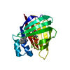
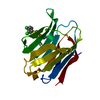
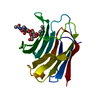
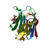
- PDB-3elx: Crystal structure of apo Zebrafish Ileal Bile Acid-Binding Protein -
+ Open data

- Basic information
| Entry | Database: PDB / ID: 3elx | ||||||
|---|---|---|---|---|---|---|---|
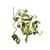
| Title | Crystal structure of apo Zebrafish Ileal Bile Acid-Binding Protein | ||||||
 Components Components | Ileal bile acid-binding protein | ||||||
 Keywords Keywords | LIPID BINDING PROTEIN / ileal bile acid-bindign protein / zebrafish / cholic acid / Lipid-binding / Transport | ||||||
| Function / homology |  Function and homology information Function and homology informationRecycling of bile acids and salts / Triglyceride catabolism / bile acid binding / fatty acid transport / fatty acid binding / nucleus / cytosol Similarity search - Function | ||||||
| Biological species |  | ||||||
| Method |  X-RAY DIFFRACTION / SYNCHROTRON / MOLECULAR REPLACEMENT / Resolution: 1.6 Å X-RAY DIFFRACTION / SYNCHROTRON / MOLECULAR REPLACEMENT / Resolution: 1.6 Å | ||||||
 Authors Authors | Capaldi, S. / Saccomani, G. / Fessas, D. / Signorelli, M. / Perduca, M. / Monaco, H.L. | ||||||
 Citation Citation | Journal: J.Mol.Biol. / Year: 2009 Title: The X-Ray structure of zebrafish (Danio rerio) ileal bile acid-binding protein reveals the presence of binding sites on the surface of the protein molecule. Authors: Capaldi, S. / Saccomani, G. / Fessas, D. / Signorelli, M. / Perduca, M. / Monaco, H.L. | ||||||
| History |
|
- Structure visualization
| Structure viewer | Molecule: MolmilJmol/JSmol |
|---|
- Downloads & links
-Download
| PDBx/mmCIF format | 3elx.cif.gz | 42.6 KB | Display | PDBx/mmCIF format |
|---|---|---|---|---|
| PDB format | pdb3elx.ent.gz | 28.6 KB | Display | PDB format |
| PDBx/mmJSON format | 3elx.json.gz | Tree view | PDBx/mmJSON format | |
| Others |  Other downloads Other downloads |
-Validation report
| Arichive directory | https://data.pdbj.org/pub/pdb/validation_reports/el/3elxftp://data.pdbj.org/pub/pdb/validation_reports/el/3elx | HTTPS FTP |
|---|
-Related structure data
| Related structure data |  3elzC  3em0C  2qo4S C: citing same article ( S: Starting model for refinement |
|---|---|
| Similar structure data |


-Links
 PDBj
PDBj

- Assembly
| Deposited unit | 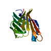
| ||||||||
|---|---|---|---|---|---|---|---|---|---|
| 1 |
| ||||||||
| Unit cell |
| ||||||||
| Details | The biological unit is the monomer. |
-Components
| #1: Protein | Mass: 15191.230 Da / Num. of mol.: 1 Source method: isolated from a genetically manipulated source Source: (gene. exp.)  |
|---|---|
| #2: Chemical | ChemComp-EDO /   Mass: 62.068 Da / Num. of mol.: 1 / Source method: obtained synthetically / Formula: C2H6O2 Mass: 62.068 Da / Num. of mol.: 1 / Source method: obtained synthetically / Formula: C2H6O2 |
| #3: Water | ChemComp-HOH /  Mass: 18.015 Da / Num. of mol.: 133 / Source method: isolated from a natural source / Formula: H2O Mass: 18.015 Da / Num. of mol.: 133 / Source method: isolated from a natural source / Formula: H2O |
-Experimental details
-Experiment
| Experiment | Method: X-RAY DIFFRACTION / Number of used crystals: 1 |
|---|
- Sample preparation
| Crystal | Density Matthews: 2.33 Å3/Da / Density % sol: 47.14 % |
|---|---|
| Crystal grow | Temperature: 293 K / Method: vapor diffusion, sitting drop / pH: 7.5 Details: 0.1 M Sodium HEPES, 1.4 M tri-sodium citrate dihydrate, 1% ethylene glycol, pH 7.5, VAPOR DIFFUSION, SITTING DROP, temperature 293K |
-Data collection
| Diffraction | Mean temperature: 100 K |
|---|---|
| Diffraction source | Source: SYNCHROTRON / Site: ESRF  / Beamline: ID29 / Wavelength: 0.97 Å / Beamline: ID29 / Wavelength: 0.97 Å |
| Detector | Type: ADSC QUANTUM 315 / Detector: CCD / Date: Dec 7, 2007 / Details: toroidal mirror |
| Radiation | Monochromator: Si 311 channel / Protocol: SINGLE WAVELENGTH / Monochromatic (M) / Laue (L): M / Scattering type: x-ray |
| Radiation wavelength | Wavelength: 0.97 Å / Relative weight: 1 |
| Reflection | Resolution: 1.6→41 Å / Num. all: 18986 / Num. obs: 18986 / % possible obs: 99.1 % / Observed criterion σ(F): 0 / Observed criterion σ(I): 0 / Redundancy: 5.2 % / Biso Wilson estimate: 15.8 Å2 / Rmerge(I) obs: 0.069 / Rsym value: 0.069 / Net I/σ(I): 16.9 |
| Reflection shell | Resolution: 1.6→1.69 Å / Redundancy: 5.3 % / Rmerge(I) obs: 0.281 / Mean I/σ(I) obs: 6.4 / Num. unique all: 2731 / Rsym value: 0.281 / % possible all: 100 |
- Processing
| Software |
| ||||||||||||||||||||||||||||||||||||||||||||||||||||||||||||||||||||||||||||||||||||||||||
|---|---|---|---|---|---|---|---|---|---|---|---|---|---|---|---|---|---|---|---|---|---|---|---|---|---|---|---|---|---|---|---|---|---|---|---|---|---|---|---|---|---|---|---|---|---|---|---|---|---|---|---|---|---|---|---|---|---|---|---|---|---|---|---|---|---|---|---|---|---|---|---|---|---|---|---|---|---|---|---|---|---|---|---|---|---|---|---|---|---|---|---|
| Refinement | Method to determine structure: MOLECULAR REPLACEMENT Starting model: 2QO4 Resolution: 1.6→30 Å / Cor.coef. Fo:Fc: 0.941 / Cor.coef. Fo:Fc free: 0.921 / SU B: 3.193 / SU ML: 0.059 / TLS residual ADP flag: LIKELY RESIDUAL / Cross valid method: THROUGHOUT / σ(F): 0 / σ(I): 0 / ESU R: 0.098 / ESU R Free: 0.1 / Stereochemistry target values: MAXIMUM LIKELIHOOD / Details: HYDROGENS HAVE BEEN ADDED IN THE RIDING POSITIONS
| ||||||||||||||||||||||||||||||||||||||||||||||||||||||||||||||||||||||||||||||||||||||||||
| Solvent computation | Ion probe radii: 0.8 Å / Shrinkage radii: 0.8 Å / VDW probe radii: 1.2 Å / Solvent model: MASK | ||||||||||||||||||||||||||||||||||||||||||||||||||||||||||||||||||||||||||||||||||||||||||
| Displacement parameters | Biso mean: 17.547 Å2
| ||||||||||||||||||||||||||||||||||||||||||||||||||||||||||||||||||||||||||||||||||||||||||
| Refinement step | Cycle: LAST / Resolution: 1.6→30 Å
| ||||||||||||||||||||||||||||||||||||||||||||||||||||||||||||||||||||||||||||||||||||||||||
| Refine LS restraints |
| ||||||||||||||||||||||||||||||||||||||||||||||||||||||||||||||||||||||||||||||||||||||||||
| LS refinement shell | Resolution: 1.6→1.641 Å / Total num. of bins used: 20
| ||||||||||||||||||||||||||||||||||||||||||||||||||||||||||||||||||||||||||||||||||||||||||
| Refinement TLS params. | Method: refined / Origin x: 27.3197 Å / Origin y: -15.2298 Å / Origin z: -8.6085 Å
|
