 Movie
Movie Controller

[English] 日本語
 Yorodumi
- PDB-2qo4: Crystal structure of zebrafish liver bile acid-binding protein co... -
+ Open data

- Basic information
| Entry | Database: PDB / ID: 2qo4 | ||||||
|---|---|---|---|---|---|---|---|
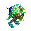
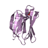
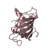
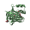
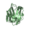
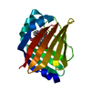
| Title | Crystal structure of zebrafish liver bile acid-binding protein complexed with cholic acid | ||||||
 Components Components | Liver-basic fatty acid binding protein | ||||||
 Keywords Keywords | LIPID BINDING PROTEIN / liver bile acid-binding protein / BABP / fatty acid-binding protein / FABP / liver (basic) fatty acid-binding protein / cholic acid / cholate / bile acid / Lipid-binding / Transport | ||||||
| Function / homology |  Function and homology information Function and homology informationbile acid binding / fatty acid transport / fatty acid binding / nucleus / cytosol Similarity search - Function | ||||||
| Biological species |  | ||||||
| Method |  X-RAY DIFFRACTION / SYNCHROTRON / MOLECULAR REPLACEMENT / Resolution: 1.5 Å X-RAY DIFFRACTION / SYNCHROTRON / MOLECULAR REPLACEMENT / Resolution: 1.5 Å | ||||||
 Authors Authors | Capaldi, S. / Saccomani, G. / Perduca, M. / Monaco, H.L. | ||||||
 Citation Citation | Journal: J.Biol.Chem. / Year: 2007 Title: A Single Amino Acid Mutation in Zebrafish (Danio rerio) Liver Bile Acid-binding Protein Can Change the Stoichiometry of Ligand Binding. Authors: Capaldi, S. / Guariento, M. / Saccomani, G. / Fessas, D. / Perduca, M. / Monaco, H.L. | ||||||
| History |
|
- Structure visualization
| Structure viewer | Molecule: MolmilJmol/JSmol |
|---|
- Downloads & links
-Download
| PDBx/mmCIF format | 2qo4.cif.gz | 42.4 KB | Display | PDBx/mmCIF format |
|---|---|---|---|---|
| PDB format | pdb2qo4.ent.gz | 27.9 KB | Display | PDB format |
| PDBx/mmJSON format | 2qo4.json.gz | Tree view | PDBx/mmJSON format | |
| Others |  Other downloads Other downloads |
-Validation report
| Arichive directory | https://data.pdbj.org/pub/pdb/validation_reports/qo/2qo4ftp://data.pdbj.org/pub/pdb/validation_reports/qo/2qo4 | HTTPS FTP |
|---|
-Related structure data
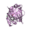
| Related structure data |  2qo5C  2qo6C  1tw4S S: Starting model for refinement C: citing same article ( |
|---|---|
| Similar structure data |


-Links
 PDBj
PDBj


- Assembly
| Deposited unit | 
| ||||||||
|---|---|---|---|---|---|---|---|---|---|
| 1 |
| ||||||||
| Unit cell |
|
-Components
| #1: Protein | Mass: 14024.225 Da / Num. of mol.: 1 Source method: isolated from a genetically manipulated source Source: (gene. exp.)  | ||||||
|---|---|---|---|---|---|---|---|
| #2: Chemical | ChemComp-CHD / 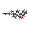  Mass: 408.571 Da / Num. of mol.: 1 / Source method: obtained synthetically / Formula: C24H40O5 Mass: 408.571 Da / Num. of mol.: 1 / Source method: obtained synthetically / Formula: C24H40O5 | ||||||
| #3: Chemical |   Mass: 60.095 Da / Num. of mol.: 3 / Source method: obtained synthetically / Formula: C3H8O Mass: 60.095 Da / Num. of mol.: 3 / Source method: obtained synthetically / Formula: C3H8O#4: Chemical | ChemComp-GOL / |   Mass: 92.094 Da / Num. of mol.: 1 / Source method: obtained synthetically / Formula: C3H8O3 Mass: 92.094 Da / Num. of mol.: 1 / Source method: obtained synthetically / Formula: C3H8O3#5: Water | ChemComp-HOH / |  Mass: 18.015 Da / Num. of mol.: 135 / Source method: isolated from a natural source / Formula: H2O Mass: 18.015 Da / Num. of mol.: 135 / Source method: isolated from a natural source / Formula: H2OHas protein modification | Y | |
-Experimental details
-Experiment
| Experiment | Method: X-RAY DIFFRACTION / Number of used crystals: 1 |
|---|
- Sample preparation
| Crystal | Density Matthews: 2.61 Å3/Da / Density % sol: 52.91 % |
|---|---|
| Crystal grow | Temperature: 298 K / Method: vapor diffusion, hanging drop / pH: 5.6 Details: 20% PEG 4000, 20% isopropanol, 0.1M sodium citrate, pH 5.6, VAPOR DIFFUSION, HANGING DROP, temperature 298.0K |
-Data collection
| Diffraction | Mean temperature: 100 K |
|---|---|
| Diffraction source | Source: SYNCHROTRON / Site: ESRF  / Beamline: ID29 / Wavelength: 0.98 Å / Beamline: ID29 / Wavelength: 0.98 Å |
| Detector | Type: ADSC QUANTUM 315 / Detector: CCD / Date: Jul 12, 2005 / Details: toroidal mirror |
| Radiation | Monochromator: toroidal mirror / Protocol: SINGLE WAVELENGTH / Monochromatic (M) / Laue (L): M / Scattering type: x-ray |
| Radiation wavelength | Wavelength: 0.98 Å / Relative weight: 1 |
| Reflection | Resolution: 1.5→33.8 Å / Num. all: 21471 / Num. obs: 21471 / % possible obs: 94.3 % / Observed criterion σ(F): 0 / Observed criterion σ(I): 0 / Redundancy: 2.8 % / Biso Wilson estimate: 10.6 Å2 / Rmerge(I) obs: 0.072 / Rsym value: 0.072 / Net I/σ(I): 13.8 |
| Reflection shell | Resolution: 1.5→1.58 Å / Redundancy: 1.9 % / Rmerge(I) obs: 0.083 / Mean I/σ(I) obs: 6.3 / Num. unique all: 2397 / Rsym value: 0.083 / % possible all: 72.6 |
- Processing
| Software |
| ||||||||||||||||||||||||||||||||||||||||||||||||||||||||||||||||||||||||||||||||||||||||||
|---|---|---|---|---|---|---|---|---|---|---|---|---|---|---|---|---|---|---|---|---|---|---|---|---|---|---|---|---|---|---|---|---|---|---|---|---|---|---|---|---|---|---|---|---|---|---|---|---|---|---|---|---|---|---|---|---|---|---|---|---|---|---|---|---|---|---|---|---|---|---|---|---|---|---|---|---|---|---|---|---|---|---|---|---|---|---|---|---|---|---|---|
| Refinement | Method to determine structure: MOLECULAR REPLACEMENT Starting model: PDB ENTRY 1TW4 Resolution: 1.5→25 Å / Cor.coef. Fo:Fc: 0.94 / Cor.coef. Fo:Fc free: 0.929 / SU B: 1.043 / SU ML: 0.041 / Isotropic thermal model: Isotropic / Cross valid method: THROUGHOUT / σ(F): 0 / σ(I): 0 / ESU R: 0.079 / ESU R Free: 0.077 / Stereochemistry target values: MAXIMUM LIKELIHOOD / Details: HYDROGENS HAVE BEEN ADDED IN THE RIDING POSITIONS
| ||||||||||||||||||||||||||||||||||||||||||||||||||||||||||||||||||||||||||||||||||||||||||
| Solvent computation | Ion probe radii: 0.8 Å / Shrinkage radii: 0.8 Å / VDW probe radii: 1.4 Å / Solvent model: MASK | ||||||||||||||||||||||||||||||||||||||||||||||||||||||||||||||||||||||||||||||||||||||||||
| Displacement parameters | Biso mean: 10.209 Å2
| ||||||||||||||||||||||||||||||||||||||||||||||||||||||||||||||||||||||||||||||||||||||||||
| Refinement step | Cycle: LAST / Resolution: 1.5→25 Å
| ||||||||||||||||||||||||||||||||||||||||||||||||||||||||||||||||||||||||||||||||||||||||||
| Refine LS restraints |
| ||||||||||||||||||||||||||||||||||||||||||||||||||||||||||||||||||||||||||||||||||||||||||
| LS refinement shell | Resolution: 1.5→1.539 Å / Total num. of bins used: 20
|
